. 2022 Nov;43(16):5081-5090.
doi: 10.1002/hbm.26080. Epub 2022 Sep 22.

Proprioceptive contribution to oculomotor control in humans

Affiliations

Proprioceptive contribution to oculomotor control in humans

Daniela Balslev et al. Hum Brain Mapp. 2022 Nov.

Abstract

Stretch receptors in the extraocular muscles (EOMs) inform the central nervous system about the rotation of one's own eyes in the orbits. Whereas fine control of the skeletal muscles hinges critically on proprioceptive feedback, the role of proprioception in oculomotor control remains unclear. Human behavioural studies provide evidence for EOM proprioception in oculomotor control, however, behavioural and electrophysiological studies in the macaque do not. Unlike macaques, humans possess numerous muscle spindles in their EOMs. To find out whether the human oculomotor nuclei respond to proprioceptive feedback we used functional magnetic resonance imaging (fMRI). With their eyes closed, participants placed their right index finger on the eyelid at the outer corner of the right eye. When prompted by a sound, they pushed the eyeball gently and briefly towards the nose. Control conditions separated out motor and tactile task components. The stretch of the right lateral rectus muscle was associated with activation of the left oculomotor nucleus and subthreshold activation of the left abducens nucleus. Because these nuclei control the horizontal movements of the left eye, we hypothesized that proprioceptive stimulation of the right EOM triggered left eye movement. To test this, we followed up with an eye-tracking experiment in complete darkness using the same behavioural task as in the fMRI study. The left eye moved actively in the direction of the passive displacement of the right eye, albeit with a smaller amplitude. Eye tracking corroborated neuroimaging findings to suggest a proprioceptive contribution to ocular alignment.

Keywords: extraocular muscles; eye; human; oculomotor; proprioceptive; visual.


Conflict of interest statement

The authors declare no conflict of interest.

Figures

FIGURE 1
FIGURE 1
Brainstem areas where the neural activity increased in response to proprioceptive stimuli. Statistical parametric map for the conjunction (passive–rest) AND (active–rest) masked exclusively with (touch–rest) is visualised for the whole brainstem above the voxel‐level threshold p < .001, uncorrected for multiple comparisons (blue). To illustrate the specificity of these results for the left side of the brainstem, the threshold for visualisation here was lower than the threshold for statistical significance for the activation peaks listed in Table 1 (p < .05 corrected for multiple comparisons). Areas that responded to both active eye movements and passive EOMs stretch (blue) are overlaid on those that were significantly activated by active eye movements alone (red, visualised for the whole brainstem above the threshold of p < .05 corrected for multiple comparisons using family‐wise error for the entire brainstem). For anatomical localisation, statistical parametric maps are overlaid on the MNI152 (ICBM) template. Activity peaks are shown in three orthogonal projections: Sagittal (top left), coronal (top right) and transversal (bottom) at the level of the (a) superior colliculus, (b) oculomotor nucleus and (c) abducens nucleus. The crosshair in each panel indicates these nuclei
FIGURE 2
FIGURE 2
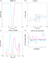
Eye movement in darkness during a brief push of the right eye leftwards. The push caused a transient leftward movement of the right eye followed by a rebound. The net active movement of the left eye mirrored the passive movement of the right eye. (a) Eye trace from one trial (participant 9) illustrating the passive movement of the right eye (cyan: Push; magenta: Rebound). (b) Eye trace of the left eye during the same trial. The colours indicate the active movements of the left eye during the two phases of the right eye displacement. (c) Detail of the left eye trace after removing any linear trend in the data. The red arrows show the net movement of the left eye during the push (cyan) and the rebound (magenta) phases of the passive right eye displacement. (d) Group data showing the net active movement of the left eye in the two phases of the passive displacement of the right eye in each individual participant. The error bars show ±1 standard error of the mean across trials calculated for each participant. Further examples of individual eye traces from participants 1–8 and 10–15 are available as Figures S3–S16

References

    1. Balslev, D. (2022). Proprioceptive contribution to oculomotor control in humans [dataset]. University of St Andrews Research Portal. 10.17630/9e107884-df5a-4de6-8327-ba809d1b2168 - DOI - PMC - PubMed
    1. Balslev, D. , Albert, N. B. , & Miall, C. (2011). Eye muscle proprioception is represented bilaterally in the sensorimotor cortex. Human Brain Mapping, 32(4), 624–631. 10.1002/hbm.21050 - DOI - PMC - PubMed
    1. Balslev, D. , & Miall, R. C. (2008). Eye position representation in human anterior parietal cortex. Journal of Neuroscience, 28, 8968–8972. - PMC - PubMed
    1. Balslev, D. , Newman, W. , & Knox, P. C. (2012). Extraocular muscle afferent signals modulate visual attention. Investigative Ophthalmology and Visual Science, 53(11), 7004–7009. 10.1167/iovs.12-10249 - DOI - PMC - PubMed
    1. Beissner, F. (2015). Functional MRI of the brainstem: Common problems and their solutions. Clinical Neuroradiology, 25(S2), 251–257. 10.1007/s00062-015-0404-0 - DOI - PubMed

Publication types