. 2022 Sep 5;12(9):859.
doi: 10.3390/membranes12090859.

Biomechanical Role of Epsin in Influenza A Virus Entry

Affiliations

Biomechanical Role of Epsin in Influenza A Virus Entry

Jophin G Joseph et al. Membranes (Basel). .

Abstract

Influenza A virus (IAV) utilizes clathrin-mediated endocytosis for cellular entry. Membrane-bending protein epsin is a cargo-specific adaptor for IAV entry. Epsin interacts with ubiquitinated surface receptors bound to IAVs via its ubiquitin interacting motifs (UIMs). Recently, epsin was shown to have membrane tension sensitivity via its amphiphilic H0 helix. We hypothesize this feature is important as IAV membrane binding would bend the membrane and clinical isolates of IAVs contain filamentous IAVs that may involve more membrane bending. However, it is not known if IAV internalization might also depend on epsin's H0 helix. We found that CALM, a structurally similar protein to epsin lacking UIMs shows weaker recruitment to IAV-containing clathrin-coated structures (CCSs) compared to epsin. Removal of the ENTH domain of epsin containing the N-terminus H0 helix, which detects changes in membrane curvature and membrane tension, or mutations in the ENTH domain preventing the formation of H0 helix reduce the ability of epsin to be recruited to IAV-containing CCSs, thereby reducing the internalization of spherical IAVs. However, internalization of IAVs competent in filamentous particle formation is not affected by the inhibition of H0 helix formation in the ENTH domain of epsin. Together, these findings support the hypothesis that epsin plays a biomechanical role in IAV entry.

Keywords: clathrin-mediated endocytosis; epsin; influenza A virus endocytosis; live-cell imaging.


Conflict of interest statement

The authors declare no conflict of interest.

Figures
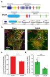
Figure 1
Figure 1
Colocalization of IAVs to ENTH/ANTH proteins containing CCSs. (a). Domain structures of epsin and CALM. GAE: gamma-adaptin ear. (b) IAVs bound to surface of RPE cells overexpressing epsin-EGFP and mCherry-Clc (left), or CALM-mCherry and EGFP-Clc (right, pseudo colored). White arrows in inset shows IAVs bound to cell surface. (c) Percentage of IAVs colocalizing with epsin and CALM to the total number of IAVs bound to the surface. The error bars denote counting error from binomial distribution. (d) Ratio of puncta intensity to background intensity of proteins colocalized with surface bound IAVs. For c and d, Ncells expressing epsin-EGFP were 15 and Ncells expressing CALM-mCherry were 10. For c and d, Ncells expressing epsin-EGFP was 15, and Ncells expressing CALM-mCherry was 10. NIAVS analyzed for epsin-EGFP and CALM was 288 and 128 respectively. For c the error bars denote standard error from binomial distribution. For d the error bar denotes standard error. *** represents p < 0.001.
Figure 2
Figure 2
Colocalization to IAVs to CCSs is disrupted in cells overexpressing epsin without a functioning ENTH domain. (ac) IAVs bound to surface of RPE cells stably expressing epsin ΔENTH EGFP, epsin mut-H0 EGFP, epsin ΔENTH ΔUIM EGFP and mCherry Clc, respectively. White arrows in inset show IAVs bound to the cell surface. (d) Fraction of IAVs colocalized with CCSs containing epsin mutants. For (d), Ncells expressing epsin-EGFP, epsin ΔENTH EGFP, epsin mut-H0 EGFP, epsin ΔENTH ΔUIM EGFP were 15, 15, 15, 15, respectively. NIAVS analyzed for epsin-EGFP, epsin ΔENTH EGFP, epsin mut-H0 EGFP, epsin ΔENTH ΔUIM EGFP were 288, 120, 147, 129, respectively. For b the error bars denote standard error from binomial distribution. The data for epsin-EGFP is reproduced from Figure 1c. The error bars denote counting error from binomial distribution. ns and *** represent not significant and p < 0.001 respectively.
Figure 3
Figure 3
Internalization of IAVs is disrupted in cells overexpressing epsin without functioning ENTH domain. (a) Flow cytometry histograms showing IAV uptake at 0 h (no infection), 1 h and 4 h in RPE cells overexpressing epsin-EGFP. (b) IAV uptake (4 h) in RPE cells with endogenous expression of epsin and overexpression of epsin WT. (c) IAV uptake (4 h) in RPE cells stably expressing epsin-EGFP, epsin ΔENTH EGFP and epsin mut-H0 EGFP. Percentage of cells with IAV internalization is shown in the inset. Experiments were repeated n = 3 and standard deviation is provided in the inset. ns represents not significant. *** represent p < 0.001.
Figure 4
Figure 4
Internalization of IAVs is disrupted by inhibition of clathrin-mediated endocytosis. (a) IAV uptake (4 h) in RPE cells with endogenous expression of epsin and overexpression of epsin WT, cells stably overexpressing epsin-EGFP and epsin mut-H0 EGFP. (b) IAV uptake (4 h) in RPE cells with endogenous expression of epsin and overexpression of epsin WT, cells stably overexpressing epsin-EGFP and epsin mut-H0 EGFP pretreated with Pitstop. (c) Percentage of cells with IAV internalization with and without Pitstop treatment. Experiments were repeated n = 3. ns represents not significant. *** represent p < 0.001.
Figure 5
Figure 5
Both spherical and filament-forming IAVs co-localize and internalize via CCSs. (a) Three example trajectories of IAV (magenta) internalization in x-y and x-z spatial orientations. Time lapse montages of WSN-WT particles co-localizing with epsin-EGFP and mCherry-Clc (top panel) and epsin mut-H0 EGFP and mCherry-Clc (bottom panel). (b) Time lapse montages of WSN-UdM particles co-localizing with epsin-EGFP and mCherry-Clc (top panel) and epsin mut-H0 EGFP and mCherry-Clc (bottom panel).
Figure 6
Figure 6
CCSs containing filament-forming IAVs strongly recruit epsin. (a) Percentage of WSN-WT and WSN-UdM particles bound to CCSs colocalizing with or without epsin or epsin mut-H0. (b) Maximum ratio of puncta intensity to background intensity of epsin or epsin mut-H0 colocalized with IAV-containing CCSs. For a and b, NIAV tracks for WSN-WT in cells expressing epsin-EGFP, WSN-WT in cells expressing epsin mut-H0 EGFP, WSN-UdM in cells expressing epsin-EGFP and WSN-UdM were 91, 105, 180, 225, respectively, and corresponding Ncells were 18, 14, 15, 21 respectively. The error bars denote standard error. ns represent not significant. *, **, and *** represent p < 0.05, p < 0.01 and p < 0.001 respectively.
Figure 7
Figure 7
Bulk uptake of WSN-UdM is not affected by mutation H0 in epsin. (a) Uptake (4 h) of WSN-WT, WSN-UdM and WSN-UdM1A by RPE cells stably expressing epsin-EGFP. Percentage of cells with IAV internalization is shown in the inset. (b) Uptake (4 h) of WSN-WT, WSN-UdM and WSN-UdM1A by RPE cells stably expressing epsin mut-H0 EGFP. Percentage of cells with IAV internalization is shown in the inset. For a and b, experiments were repeated n = 3 and standard deviation is provided in the inset. ns represents not significant. *, and *** represent p < 0.05 and p < 0.001 respectively.

References

    1. Doherty G.J., McMahon H.T. Mechanisms of Endocytosis. Annu. Rev. Biochem. 2009;78:857–902. doi: 10.1146/annurev.biochem.78.081307.110540. - DOI - PubMed
    1. Cossart P., Helenius A. Endocytosis of viruses and bacteria. Cold Spring Harb. Perspect. Biol. 2014;6:a016972. doi: 10.1101/cshperspect.a016972. - DOI - PMC - PubMed
    1. Bayati A., Kumar R., Francis V., McPherson P.S. SARS-CoV-2 infects cells after viral entry via clathrin-mediated endo-cytosis. J. Biol. Chem. J. Biol. Chem. 2021;296:100306. - PMC - PubMed
    1. Piccini L.E., Castilla V., Damonte E.B. Dengue-3 Virus Entry into Vero Cells: Role of Clathrin-Mediated Endocytosis in the Outcome of Infection. PLoS ONE. 2015;10:e0140824. doi: 10.1371/journal.pone.0140824. - DOI - PMC - PubMed
    1. van der Schaar H.M., Rust M.J., Chen C., van der Ende-Metselaar H., Wilschut J., Zhuang X., Smit J.M. Dissecting the Cell Entry Pathway of Dengue Virus by Single-Particle Tracking in Living Cells. PLOS Pathog. 2008;4:e1000244. doi: 10.1371/journal.ppat.1000244. - DOI - PMC - PubMed

LinkOut - more resources