. 2022 Aug 29;14(9):595.
doi: 10.3390/toxins14090595.

Functional and Immunological Studies Revealed a Second Superantigen Toxin in Staphylococcal Enterotoxin C Producing Staphylococcus aureus Strains

Affiliations

Functional and Immunological Studies Revealed a Second Superantigen Toxin in Staphylococcal Enterotoxin C Producing Staphylococcus aureus Strains

Andreas Roetzer et al. Toxins (Basel). .

Abstract

Staphylococcus aureus is a human and animal pathogen as well as a commensal bacterium. It can be a causative agent of severe, life-threatening infections with high mortality, e.g., toxic shock syndrome, septic shock, and multi-organ failure. S. aureus strains secrete a number of toxins. Exotoxins/enterotoxins are considered important in the pathogenesis of the above-mentioned conditions. Exotoxins, e.g., superantigen toxins, cause uncontrolled and polyclonal T cell activation and unregulated activation of inflammatory cytokines. Here we show the importance of genomic analysis of infectious strains in order to identify disease-causing exotoxins. Further, we show through functional analysis of superantigenic properties of staphylococcal exotoxins that even very small amounts of a putative superantigenic contaminant can have a significant mitogenic effect. The results show expression and production of two distinct staphylococcal exotoxins, SEC and SEL, in several strains from clinical isolates. Antibodies against both toxins are required to neutralise the superantigenic activity of staphylococcal supernatants and purified staphylococcal toxins.

Keywords: Staphylococcus aureus; mitogenesis; neutralisation; staphylococcal enterotoxins.


Conflict of interest statement

The authors declare no conflict of interest.

Figures

Figure 1
Figure 1
(a) Superantigen toxins have a broad range of MNC activation. Polyclonal T cell stimulation can be detected with toxin concentrations as low as 10 pg/mL. Proliferation of human peripheral blood MNCs stimulated with purified supernatant from S. aureus 19095 (SUP) and wild-type recombinant SEC and SEL. Incorporation of [3H] thymidine was counted. Experiments were carried out with MNCs from two different donors. Wild-type recombinant and native proteins were diluted in RPMI 1640 complete medium. MNCs were adjusted to 1 × 10⁶ cells per mL. Phytohaemagglutinin (PHA) was used as a positive control. (b) Estimation of SEC concentrations in supernatants of staphylococcal strain 19095. Well-defined amounts of recombinant SEC were compared to different amounts of supernatant of strain 19095. Anti-SEC antiserum was used for detection. (c) SEC and SEL are present in different concentrations in the supernatant of staphylococcal strain 19095. Estimation of SEL concentrations in purified supernatants of staphylococcal strain 19095, analysed by Western blot. An amount of 50 ng of bacterial supernatant of laboratory strain 19095 (SUP) or recombinant wild-type proteins (SEC, SEL, SElM, SElN) was loaded on a polyacrylamide gel, electrophoresed, and transferred to a nitrocellulose membrane. PageRulerTM Plus (Biorad; left panel) and Precision Plus ProteinTM (Biorad; right panel) were added as ladder to identify SEL bands. Anti-SEL antiserum was used for detection of SEL.
Figure 2
Figure 2
Anti-SEC antiserum alone cannot completely neutralise proliferation of MNCs induced by supernatant of staphylococcal laboratory strain 19095. Neutralisation of proliferation induced by recombinant SEC (left panel) or supernatants of staphylococcal strain 19095 (staph sup (SUP), right panel) is shown. Bars from antisera against purified staphylococcal supernatant are shown in red, bars from antisera against recombinant superantigen are shown in yellow, and bars of controls (no addition of sera, and MNCs only) are given in green. Proliferation of human peripheral blood MNCs was quantified by measuring [3H] thymidine incorporated. Experiments were conducted with MNCs from two independent donors. MNCs were adjusted to 1 × 10⁶ cells per mL. Percentage of neutralisation results (lower panels) were taken from means of triplicate values (tested in two donors) and normalised against the negative control (MNC).
Figure 3
Figure 3
Both SEC and SEL antisera are required to neutralise proliferation of MNCs induced by the supernatant of staphylococcal laboratory strain 19095. Bars from antisera against recombinant superantigens SEC/SEL are shown in violet, and bars of controls are given in green (no addition of sera, PHA) and dark green (MNCs only). Proliferation of human peripheral blood MNCs was quantified by measuring [3H] thymidine incorporated. Experiments were conducted with MNCs from two independent donors. MNCs were adjusted to 1 × 10⁶ cells per mL. Percentage of neutralisation results (lower panels) were taken from means of triplicate values (tested in two donors) and normalised against the negative control (MNC).
Figure 4
Figure 4
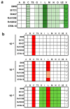
(a) Genomic superantigen repertoire of the six strains reported in this study. Sequencing results revealed that the six strains expressing SEC and SEL also contain the genes for the egc operon (seg, sei, selm, seln, selo, and selu2, lined boxes). Big letters indicate (prominent) superantigen genes tested (e.g., A = sea, X = selx). Selu2 was the only version of selu genes present. Quotation marks indicate truncated version of selx (boxes shown in green; members of the egc operon are shown with same stripe pattern). Blood strains are indicated as B, colonising strains are indicated as Rv or N-10. (b) Neutralisation of superantigen containing supernatants of six different strains at different dilutions. Supernatant diluted 10−5 needs antisera against SEC and SEL to be neutralised (red boxes), although strains Rv51398 and Rv52832 show only weak proliferation without anti-SEL antiserum (rosy boxes, middle panel). At a supernatant dilution of 10−4, proliferation is only inhibited by adding antisera against SEC as well as SEL, G, I, M, N, and O (lower panel).
Figure 5
Figure 5
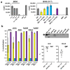
Proliferation of human peripheral blood mononuclear cells is stimulated by supernatants comprising the two superantigens SEC and SEL and can be neutralised by antisera against both. (a) Induced proliferation was measured by incorporation of [3H] thymidine. Each experiment was performed in triplicate. Bacterial strains were centrifuged and filtrated, cell-free supernatants including sub-cellular particles were analysed. Diluted supernatants were neutralised with antisera (x-axis annotation; bars for SEC neutralization are shown in yellow, SEL in blue, and SEC/SEL in violet) for 1 h at 37 °C (900 rpm). The mitogen PHA was added to neutralisations as control to reveal cytotoxic effects. (b) Percentages of neutralisation of supernatants. Different suspensions of antisera were applied in a 1:10 dilution to specific 10−5-fold diluted supernatants of tested SEC-producing strains. After incubation, mixes were added to isolated blood cells, and incorporation of [3H] thymidine was measured. Each experiment was performed in triplicate (left panel). Western blot analysis of collected bacterial samples with a polyclonal antiserum raised against SEL. PageRulerTM Plus (Biorad) was added as ladder to identify correct bands (25 kDa and 15 kDa bands are shown on the blot). An amount of 50 ng SEL wild-type recombinant protein was used as positive control (right upper panel). Scattered dot plots with geometric means (including 95% CI) of percentages of supernatant neutralisations are shown. Three blood isolates and three colonising isolates tested in triplicates resulted in nine neutralisation values for both groups, which were included here (right lower panel).

References

    1. Mulcahy M.E., McLoughlin R.M. Host-Bacterial Crosstalk Determines Staphylococcus aureus Nasal Colonization. Trends Microbiol. 2016;24:872–886. doi: 10.1016/j.tim.2016.06.012. - DOI - PubMed
    1. Laux C., Peschel A., Krismer B. Staphylococcus aureus Colonization of the Human Nose and Interaction with Other Microbiome Members. Microbiol. Spectr. 2019;7:34. doi: 10.1128/microbiolspec.GPP3-0029-2018. - DOI - PMC - PubMed
    1. Wertheim H.F., Melles D.C., Vos M.C., van Leeuwen W., van Belkum A., Verbrugh H.A., Nouwen J.L. The role of nasal carriage in Staphylococcus aureus infections. Lancet Infect. Dis. 2005;5:751–762. doi: 10.1016/S1473-3099(05)70295-4. - DOI - PubMed
    1. Tong S.Y., Davis J.S., Eichenberger E., Holland T.L., Fowler V.G., Jr. Staphylococcus aureus infections: Epidemiology, pathophysiology, clinical manifestations, and management. Clin. Microbiol. Rev. 2015;28:603–661. doi: 10.1128/CMR.00134-14. - DOI - PMC - PubMed
    1. Lowy F.D. Staphylococcus aureus infections. N. Engl. J. Med. 1998;339:520–532. doi: 10.1056/NEJM199808203390806. - DOI - PubMed

Publication types