. 2022 Sep 1;14(9):605.
doi: 10.3390/toxins14090605.

Arabic Gum Could Alleviate the Aflatoxin B1-provoked Hepatic Injury in Rat: The Involvement of Oxidative Stress, Inflammatory, and Apoptotic Pathways

Affiliations

Arabic Gum Could Alleviate the Aflatoxin B1-provoked Hepatic Injury in Rat: The Involvement of Oxidative Stress, Inflammatory, and Apoptotic Pathways

Noha Ahmed et al. Toxins (Basel). .

Abstract

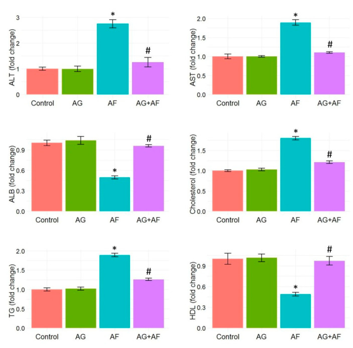
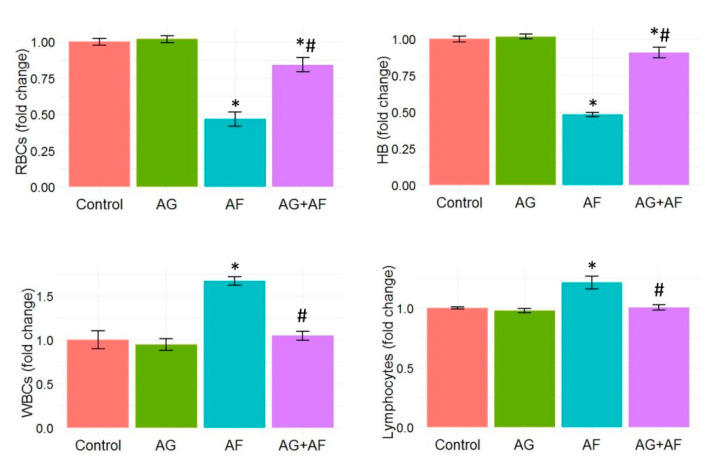
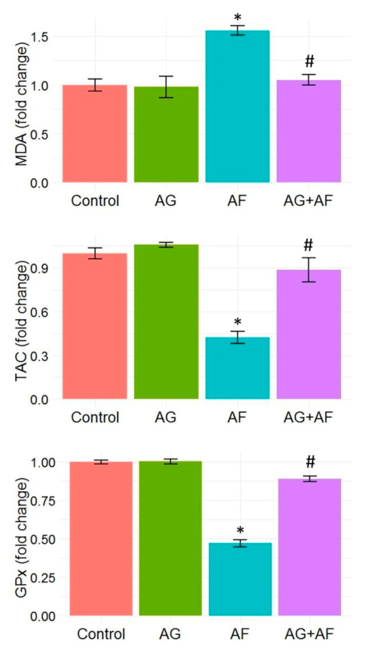
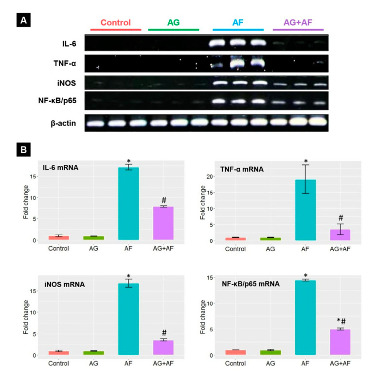
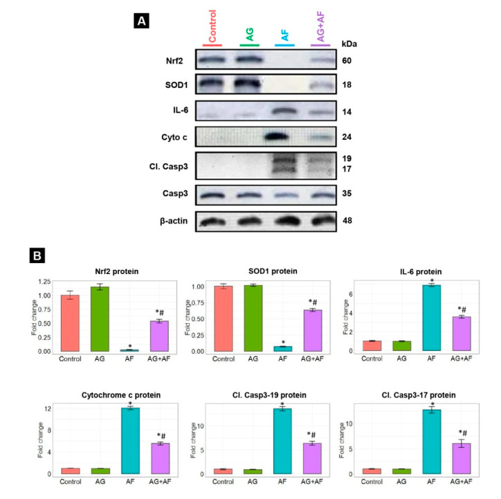
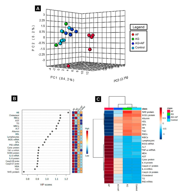
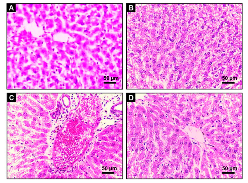
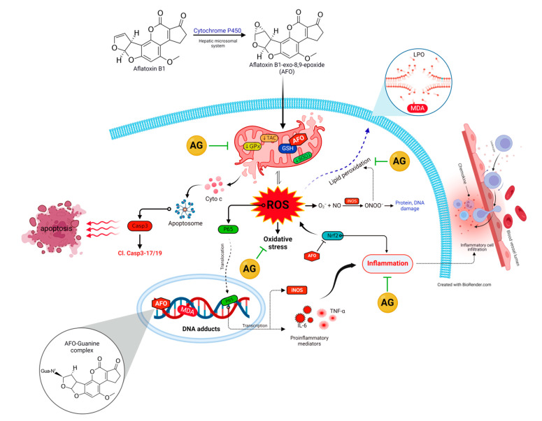
Aflatoxin B1 (AF) is an unavoidable environmental pollutant that contaminates food, feed, and grains, which seriously threatens human and animal health. Arabic gum (AG) has recently evoked much attention owing to its promising therapeutic potential. Thus, the current study was conducted to look into the possible mechanisms beyond the ameliorative activity of AG against AF-inflicted hepatic injury. Male Wistar rats were assigned into four groups: Control, AG (7.5 g/kg b.w/day, orally), AF (200 µg/kg b.w), and AG plus AF group. AF induced marked liver damage expounded by considerable changes in biochemical profile and histological architecture. The oxidative stress stimulated by AF boosted the production of plasma malondialdehyde (MDA) level along with decreases in the total antioxidant capacity (TAC) level and glutathione peroxidase (GPx) activity. Additionally, AF exposure was associated with down-regulation of the nuclear factor erythroid2-related factor2 (Nrf2) and superoxide dismutase1 (SOD1) protein expression in liver tissue. Apoptotic cascade has also been evoked following AF-exposure, as depicted in overexpression of cytochrome c (Cyto c), cleaved Caspase3 (Cl. Casp3), along with enhanced up-regulation of inflammatory mediators such as tumor necrosis factor-α (TNF-α), interleukin (IL)-6, inducible nitric oxide synthase (iNOS), and nuclear factor kappa-B transcription factor/p65 (NF-κB/p65) mRNA expression levels. Interestingly, the antioxidant and anti-inflammatory contents of AG may reverse the induced oxidative damage, inflammation, and apoptosis in AF-exposed animals.

Keywords: Arabic gum; aflatoxin B1; apoptosis; inflammatory cytokines; liver injury; oxidative stress.


Conflict of interest statement

The authors declare no conflict of interest.

Figures

Figure 1
Figure 1
Bar plot panel of liver biochemical parameters and lipid profile upon AF and/or AG treatment. Values shown are mean ± SE (n = 5). ALB, albumin; ALT, alanine aminotransferase; AST, aspartate aminotransferase; HDL, high-density lipoprotein; TG, triglyceride. p < 0.01; * AF vs control; # AG+AF vs AF.
Figure 2
Figure 2
Bar plot panel of hematological profile upon AF and/or AG treatment. Values shown are mean ± SE (n = 5). RBC, red blood cells; HB, hemoglobin; and WBCs, white blood cells. p < 0.01; * AF vs control; # AG+AF vs AF.
Figure 3
Figure 3
Bar plot panel of plasma antioxidant and peroxidation biomarkers changes upon AF and/or AG treatment. Values shown are mean ± SE (n = 5). GPx, glutathione peroxidase, MDA, malondialdehyde; TAC, total antioxidant capacity. p < 0.01; * AF vs control; # AG+AF vs AF.
Figure 4
Figure 4
The mRNA expression of proinflammatory cytokines in liver upon AF and/or AG treatment. (A) representative bands for IL-6, TNF-α, iNOS, NF-κB/p65, and β-actin genes; (B) Bar plot panel of the semiquantitative analysis of mRNA levels of proinflammatory cytokines after normalization against β-actin. p < 0.01; * AF vs control; # AG+AF vs AF.
Figure 5
Figure 5
Protein expression of inflammatory cytokine, antioxidant, and apoptotic proteins in liver upon AF and/or AG treatment. (A) Typical immunoblots for IL-6, Nrf2, SOD1, Cyto c, Cl. Casp3-17/19, Casp3, and β-actin proteins; (B) Bar plot panel for semiquantitative data were created from immunoblot after normalization against β-actin. p < 0.01; * AF vs control; # AG+AF vs AF.
Figure 6
Figure 6
Principal components analysis (PCA) and data clustering analysis of AG versus AF-induced liver toxicity. (A) 3D score plot of PCA for discerning the four experimental groups (Control, AG, AF, and AG+ AF). Percentage values specified on the axes depict the contribution rate of PC1 (84.2%), PC2 (6.2%), and PC3 (2.7%) to the overall number of variations; (B) variable importance in projection (VIP): the colored boxes on the right display the relative concentrations of the relevant measured parameters in each study group, while, the contribution intensity is indicated by a colored scale spanning from the highest (red) to lowest (blue); (C) hierarchical clustering heatmap enables intuitive visualization of all data sets. Each colored cell on the map denotes the concentration values, with variable averages in rows and different treatment sets in columns. Dark red is the highest value on the gradation scale, and blue represents the lowest value.
Figure 7
Figure 7
Histopathology of liver tissue in Control, AG, AF, and AG+ AF-treated groups. Grossly normal architecture of hepatic lobules was observed in the Control (A), and AG-treated (B) rats; AF (C) liver section of AF-treated group showed periportal cytoplasmic vacuolation with fatty degeneration, severe hemorrhage, bile duct hyperplasia, and inflammatory cellular infiltration; AG+ AF-treated group (D) exhibited substantial improvement in hepatic architecture, indicated by mild fatty vacuolation and portal inflammation.
Figure 8
Figure 8
The molecular insights behind the protective effect of AG following AF-induced toxicity. AFO, aflatoxin-exo-8,9-epoxide; AG, Arabic gum; Cl. Casp3-17/19, cleaved Caspase3-17/19; Cyto c, cytochrome c; GPx, glutathione peroxidase; GSH, reduced-glutathione; IL-6, interleukin-6; iNOS, inducible nitric oxide synthase; LPO, lipid peroxidation; MDA, malondialdehyde; NF-κB/p65, nuclear factor kappa-B transcription factor/p65; NO, nitric oxide; Nrf2, nuclear factor erythroid2–related factor2; ONOO, toxic peroxy-nitrite species; ROS, reactive oxygen species; SOD1, superoxide dismutase1; TAC, total antioxidant capacity; TNF-α, tumor necrosis factor-α.

Similar articles

Cited by

References

    1. Zhu F., Zhao X., Li J., Guo L., Bai L., Qi X. A new compound Trichomicin exerts antitumor activity through STAT3 signaling inhibition. Biomed. Pharmacother. 2020;121:109608. doi: 10.1016/j.biopha.2019.109608. - DOI - PubMed
    1. Zhang M., Jiao P., Wang X., Sun Y., Liang G., Xie X., Zhang Y. Evaluation of Growth Performance, Nitrogen Balance and Blood Metabolites of Mutton Sheep Fed an Ammonia-Treated Aflatoxin B1-Contaminated Diet. Toxins. 2022;14:361. doi: 10.3390/toxins14050361. - DOI - PMC - PubMed
    1. COMMISSION REGULATION (EC) No 401/2006 Laying down the methods of sampling and analysis for the official control of the levels of mycotoxins in foodstuffs. Comm. Regul. 2006;70:12.
    1. Abdel-Daim M.M., Abdeen A., Jalouli M., Abdelkader A., Megahed A., Alkahtane A., Almeer R., Alhoshani N.M., Al-Johani N.S., Alkahtani S., et al. Fucoidan supplementation modulates hepato-renal oxidative stress and DNA damage induced by aflatoxin B1 intoxication in rats. Sci. Total Environ. 2021;768:144781. doi: 10.1016/j.scitotenv.2020.144781. - DOI - PubMed
    1. Yilmaz S., Bag H. Aflatoxin B1: Mechanism, oxidative stress, and effects on animal health. Insights Vet. Sci. 2022;6:17–24. doi: 10.29328/journal.ivs.1001037. - DOI

Publication types

MeSH terms