. 2022 Sep 19;5(5):71.
doi: 10.3390/mps5050071.

Single and Multiplex Immunohistochemistry to Detect Platelets and Neutrophils in Rat and Porcine Tissues

Affiliations

Single and Multiplex Immunohistochemistry to Detect Platelets and Neutrophils in Rat and Porcine Tissues

Stephanie Arnold et al. Methods Protoc. .

Abstract

Platelet-neutrophil complexes (PNCs) occur during the inflammatory response to trauma and infections, and their interactions enable cell activation that can lead to tissue destruction. The ability to identify the accumulation and tissue localisation of PNCs is necessary to further understand their role in the organs associated with blast-induced shock wave trauma. Relevant experimental lung injury models often utilise pigs and rats, species for which immunohistochemistry protocols to detect platelets and neutrophils have yet to be established. Therefore, monoplex and multiplex immunohistochemistry protocols were established to evaluate the application of 22 commercially available antibodies to detect platelet (nine rat and five pig) and/or neutrophil (four rat and six pig) antigens identified as having potential selectivity for porcine or rat tissue, using lung and liver sections taken from models of polytrauma, including blast lung injury. Of the antibodies evaluated, one antibody was able to detect rat neutrophil elastase (on frozen and formalin-fixed paraffin embedded (FFPE) sections), and one antibody was successful in detecting rat CD61 (frozen sections only); whilst one antibody was able to detect porcine MPO (frozen and FFPE sections) and antibodies, targeting CD42b or CD49b antigens, were able to detect porcine platelets (frozen and FFPE and frozen, respectively). Staining procedures for platelet and neutrophil antigens were also successful in detecting the presence of PNCs in both rat and porcine tissue. We have, therefore, established protocols to allow for the detection of PNCs in lung and liver sections from porcine and rat models of trauma, which we anticipate should be of value to others interested in investigating these cell types in these species.

Keywords: immunohistochemistry; neutrophils; platelets; porcine; rat.


Conflict of interest statement

The authors declare no conflict of interest.

Figures

Figure 1
Figure 1
Schematic of single-stain and multiplex immunohistochemistry reporter systems. In single-stained immunohistochemistry (IHC) (above), neutrophils and platelets are detected in separate tissue sections using a horse radish peroxidase (HRP)-dependent reporting system. The 3,3’-diaminobenzidine (DAB) substrate forms a brown precipitate in positively stained regions. In multiplex IHC (below), platelets and neutrophils can be detected in the same tissue section. Platelets are detected using a HRP-dependent reaction, whilst neutrophils are detected using an alkaline phosphatase (AP) dependent reaction. Blue and red colour substrates allow for visualisation of colocalised events.
Figure 2
Figure 2
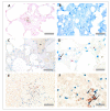
Neutrophil immunohistochemistry in rat lung and liver tissues. For protocol optimisation, sections were immunostained with primary antibody (A) and without primary antibody (B), to detect any unspecific binding of detection reagents. Representative images of neutrophil immunohistochemistry (anti-MPO) in FFPE rat lung tissue (C,D) and FFPE rat liver tissue (E,F) are also shown. Asterisks denote blood vessels; black dotted lines denote airway wall. Scale bar = 100 µm in images (A,B,D,E) (×20 objective), 50 µm in image (F) (×40 objective) and 20 µm in image (C) (×63 objective).
Figure 3
Figure 3
Platelet immunohistochemistry in rat lung and liver tissues. For protocol optimisation, sections were immunostained with primary antibody (A) and without primary antibody (B), to ensure no unspecific binding of detection reagents. Representative images of platelet immunohistochemistry (anti-CD61) in frozen rat lung tissue (C,D) and frozen rat liver tissue (E,F) are also shown. Asterisks denote blood vessels. Arrows denote CD61+ events. Scale bar = 50 µm in (A,C) (×40 objective), 100 µm in (B,D,E,F) (×20 objective).
Figure 4
Figure 4
Platelet neutrophil complex multiplex immunohistochemistry in rat lung and liver tissues. Frozen rat tissues were prepared and immunostained with anti-myeloperoxidase (MPO) antibody (ab9535, Abcam, UK) and anti-CD61 antibody (PB9647, Boster, USA), prepared at 1/25 and 1/100 dilutions, respectively. Platelets appear red, and neutrophils appear blue. Representative images of platelet neutrophil complex (PNC) staining in rat lung tissue (A,B) and rat liver tissue (C,D). Arrows denote CD61+ MPO+ events. Scale bar = 100 µm in Figures (A,B) (×20 objective) and 30 µm in Figures (B,C) (×63 objective).
Figure 5
Figure 5
Neutrophil immunohistochemistry in pig lung and liver tissues. For protocol optimisation, consecutive sections were immunostained with primary antibody (A) and without primary antibody (B), to ensure no unspecific binding of detection reagents. Representative images of neutrophil immunohistochemistry (anti-MPO) in FFPE pig lung tissue (C,D) and FFPE pig liver tissue (E,F) are also shown. Asterisks denote blood vessels. Scale bar = 100 µm in Figures (AC,E,F) (×20 objective), 20 µm in Figure (D) (×63 objective).
Figure 6
Figure 6
Platelet immunohistochemistry in frozen pig lung and liver tissues. Consecutive sections were immunostained with primary antibody (A) and without primary antibody (B), to detect unspecific binding of detection reagents. Representative images of platelet immunohistochemistry (anti-CD49b) in frozen pig lung tissue (C) are also shown. Signal is absent in the reagent only control (D). Platelets were also observed in the liver tissue (E,F). Asterisks denote blood vessels. Arrows denote CD49+ platelet events. Scale bar = 50 µm in Figures (A,E) (×40 objective), 100 µm in Figures (B,D) (×20 objective) and 20 µm in Figures (C,F) (×63 objective).
Figure 7
Figure 7
Platelet immunohistochemistry in FFPE pig lung and liver tissues. Sections were immunostained with primary antibody (A) and without primary antibody (B), to ensure no unspecific binding of detection reagents. Representative images of neutrophil immunohistochemistry (anti-CD42b) in FFPE pig lung tissue (C,D) and FFPE pig liver tissue (E,F) are also shown. Asterisks denote blood vessels. Arrows denote CD42b+ platelet events. Scale bar = 100 µm in Figures (A,D) (×20 objective), 50 µm in Figure (B,F) and 30 µm in Figures (C,E) (×63 objective).
Figure 8
Figure 8
Platelet neutrophil complex multiplex immunohistochemistry in pig lung and liver tissues. To ensure colour detection systems were functional, separate sections were immunostained with either anti-CD42b shown in red (A) or anti-MPO shown in blue. Representative images of neutrophil immunohistochemistry (anti-CD42b) in pig lung tissue (C,D) and pig liver tissue (E,F) are also shown. Asterisks denote blood vessels. Arrows point to PNCs. Scale bar = 50 µm in Figures (A,B) (×40 objective), 100 µm in Figure (C,E) (×20 objective) and 30 µm in Figures (C,E) (×63 objective) for neutrophil complex multiplex immunohistochemistry in pig lung and liver tissues. Formalin fixed paraffin wax embedded (FFPE), and pig tissues were prepared and immunostained with anti-myeloperoxidase (MPO) antibody (ab9535, Abcam, Cambridge, UK) and anti-CD42b antibody (ab183345, Abcam, Cambridge, UK), prepared at 1/50 and 1/100 dilutions, respectively. Platelets appear red, and neutrophils appear blue. To ensure colour detection systems were functional, separate sections were immunostained with either anti-CD42b shown in red (A) or anti-MPO shown in blue. Representative images of neutrophil immunohistochemistry (anti-CD42b) in pig lung tissue (C,D) and pig liver tissue (E,F) are also shown. Asterisks denote blood vessels. Arrows point to PNCs. Scale bar = 50 µm in Figures (A,B) (×40 objective), 100 µm in Figure (C,E) (×20 objective) and 30 µm in Figures (C,E) (×63 objective).

References

    1. Middleton E.A., Weyrich A.S., Zimmerman G.A. Platelets in Pulmonary Immune Responses and Inflammatory Lung Diseases. Physiol. Rev. 2016;96:1211–1259. doi: 10.1152/physrev.00038.2015. - DOI - PMC - PubMed
    1. Cleary S.J., Hobbs C., Amison R.T., Arnold S., O’Shaughnessy B.G., Lefrançais E., Mallavia B., Looney M.R., Page C.P., Pitchford S.C. LPS-induced Lung Platelet Recruitment Occurs Independently from Neutrophils, PSGL-1, and P-Selectin. Am. J. Respir. Cell Mol. Biol. 2019;61:232–243. doi: 10.1165/rcmb.2018-0182OC. - DOI - PMC - PubMed
    1. Looney M.R., Nguyen J.X., Hu Y., Van Ziffle J.A., Lowell C.A., Matthay M.A. Platelet depletion and aspirin treatment protect mice in a two-event model of transfusion-related acute lung injury. J. Clin. Investig. 2009;119:3450–3461. doi: 10.1172/JCI38432. - DOI - PMC - PubMed
    1. Chavko M., Prusaczyk W.K., McCarron R.M. Lung Injury and Recovery After Exposure to Blast Overpressure. J. Trauma. 2006;61:933–942. doi: 10.1097/01.ta.0000233742.75450.47. - DOI - PubMed
    1. Scott T., Kirkman E., Haque M., Gibb I.E., Mahoney P., Hardman J. Primary blast lung injury—A review. Br. J. Anaesth. 2017;118:311–316. doi: 10.1093/bja/aew385. - DOI - PubMed