. 2022 Sep 23;377(6613):1413-1419.
doi: 10.1126/science.abo4837. Epub 2022 Sep 22.

Quality assurance of hematopoietic stem cells by macrophages determines stem cell clonality

Affiliations

Quality assurance of hematopoietic stem cells by macrophages determines stem cell clonality

Samuel J Wattrus et al. Science. .

Abstract

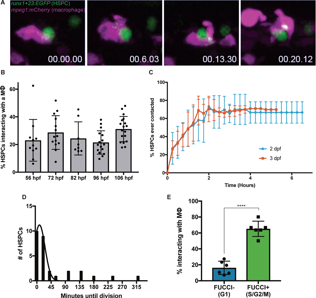
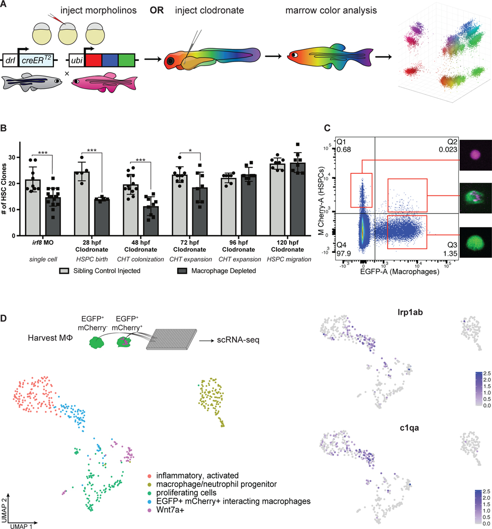
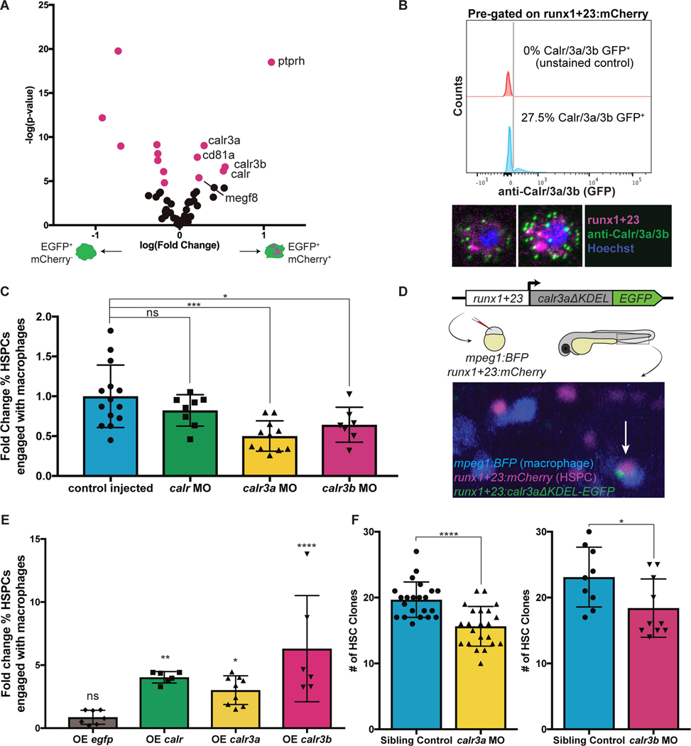
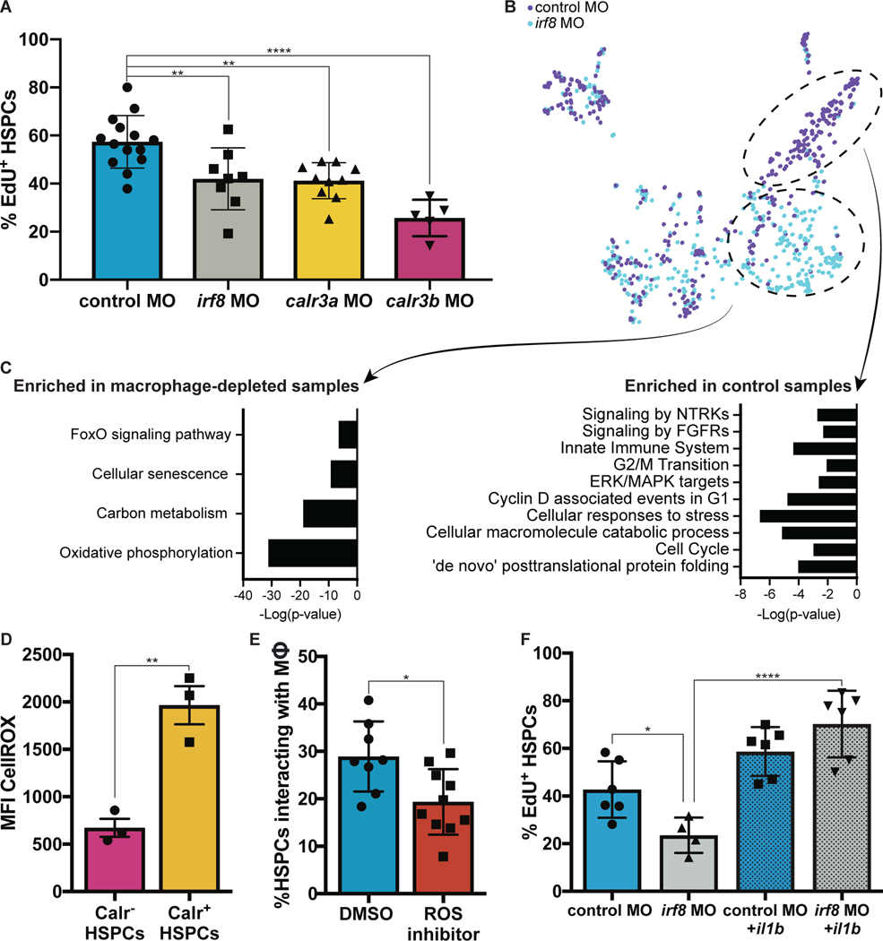
Tissue-specific stem cells persist for a lifetime and can differentiate to maintain homeostasis or transform to initiate cancer. Despite their importance, there are no described quality assurance mechanisms for newly formed stem cells. We observed intimate and specific interactions between macrophages and nascent blood stem cells in zebrafish embryos. Macrophage interactions frequently led to either removal of cytoplasmic material and stem cell division or complete engulfment and stem cell death. Stressed stem cells were marked by surface Calreticulin, which stimulated macrophage interactions. Using cellular barcoding, we found that Calreticulin knock-down or embryonic macrophage depletion reduced the number of stem cell clones that established adult hematopoiesis. Our work supports a model in which embryonic macrophages determine hematopoietic clonality by monitoring stem cell quality.


Conflict of interest statement

Competing interests: All other authors declare that they have no competing interests.

Figures

Fig. 1.
Fig. 1.. Macrophages make intimate interactions with newly formed HSPCs.
(A) Time lapse live-imaging identifies prolonged cell-cell contacts between runx1+23:EGFP+ HSPCs and mpeg1:mCherry+ primitive macrophages involving exchange of fluorescent material. (B) Approximately 20–30% of HSPCs interact with macrophages in the CHT at any one time from 56 hpf to 106 hpf. Mean +/− s.d. (C) High-resolution tracking of individual runx1+23:mCherry+ cells over several hours in the CHT reveals that the majority of HSPCs eventually make sustained contact with macrophages (> 5 minutes). Mean +/− s.d. (D) HSPCs frequently complete a cell division shortly after macrophage interactions. Approximately 81% of HSPC divisions occur within 30 minutes of a macrophage interaction. Mean +/− s.d. (E) Around 65% of Fucci+ HSPCs in S/G2/M phases interact with macrophages at any one time, as compared to less than 20% of Fucci HSPCs. Mean +/− s.d., Unpaired t test; ****P<0.0001.
Fig. 2.
Fig. 2.. Macrophages in the CHT regulate stem cell clonality.
(A) A schematic overview of the Zebrabow-M system: animals with 15–20 insertions of a multicolor fluorescent cassette are crossed to the draculin:CreERT2 line to enable clonal labeling of lateral plate mesoderm lineages. By treating with 4-OHT at 24 hpf just after HSC specification, individual stem cell lineages express unique fluorescent hues which can be quantified in the adult marrow. (B) Families of Zebrabow-M;draculin:CreERT2 animals injected with either clodronate liposomes or the irf8 morpholino exhibit reduced numbers of HSC clones in the adult marrow, even when macrophages are not depleted until after emergence from the VDA. Mean +/− s.d., Unpaired t test; *P<0.05, ***P<0.001. (C) Macrophages (mpeg1:EGFP+) which have interacted with HSPCs (runx1+23:mCherry+) and removed fluorescent material can be harvested by FACS. (D) Macrophages which engage HSPCs are marked by lrp1ab and c1qa. Spectral scale reports z-scores.
Fig. 3.
Fig. 3.. Calreticulin drives HSPC-macrophage interactions to regulate clonality.
(A) Analysis of differentially enriched potential surface proteins from interacting macrophages identifies three paralogs of Calreticulin. (B) Flow cytometry shows ~30% of runx1+23:mCherry+ HSPCs stain for surface Calreticulin. (C) Morpholino knock-down of calr3a or calr3b significantly reduces the fraction of HSPCs interacting with macrophages at any one time. Mean +/− s.d., One-way ANOVA with Dunnett’s multiple comparisons test; *P<0.05, ***P<0.001. (D) Calreticulin paralogs without the ER-retention KDEL sequence were fused to EGFP, driven by the HSPC-specific runx1+23 enhancer, and injected into stable runx1+23:mCherry;mpeg1:BFP embryos. Mosaic animals overexpress Calreticulin in a random subset of HSPCs. Arrow indicates an HSPC overexpressing calr3a engaged by a macrophage. (E) HSPCs overexpressing calr, calr3a, or calr3b are more frequently engaged by macrophages compared to non-overexpressing HSPCs in the same embryos. Overexpressing egfp alone has no effect. Mean +/− s.d., One-way ANOVA with Dunnett’s multiple comparisons test; *P<0.05, **P<0.01, ****P<0.0001. (F) Knock-down of calr3a or calr3b reduces the number of HSC clones that contribute to adult hematopoiesis. Mean +/− s.d., Unpaired t test; *P<0.05, ****P<0.0001.
Fig. 4.
Fig. 4.. Macrophages buffer HSPC stress and regulate HSPC expansion.
(A) EdU staining of runx1+23:mCherry embryos injected with either the calr3a, calr3b, or irf8 morpholinos identifies significant reduction in proliferating HSPCs at 3 dpf. Mean +/− s.d., One-way ANOVA with Dunnett’s multiple comparisons test; **P<0.01, ****P<0.0001. (B)(C) Single-cell mRNA-seq analysis of runx1+23+ FACS-purified cells from irf8 or control morphants reveals a population of stressed HSPCs that persist in the absence of macrophages and a population of cycling cells enriched in the control sample. (D) Embryonic HSPCs marked by surface Calreticulin exhibit higher levels of ROS. (E) ROS inhibition with diphenylene iodonium significantly reduces macrophage-HSPC interactions. Mean +/− s.d., Unpaired t test; *P<0.05. (F) Expression of il1b by heat shock rescues the effect of macrophage depletion on HSPC proliferation. Mean +/− s.d., One-way ANOVA with Sidak’s multiple comparisons test; *P<0.05, ****P<0.001.

Comment in

References

    1. Kissa K, Herbomel P, Blood stem cells emerge from aortic endothelium by a novel type of cell transition. Nature 464, 112–115 (2010). - PubMed
    1. Bertrand JY et al., Haematopoietic stem cells derive directly from aortic endothelium during development. Nature 464, 108–111 (2010). - PMC - PubMed
    1. Murayama E et al., Tracing hematopoietic precursor migration to successive hematopoietic organs during zebrafish development. Immunity 25, 963–975 (2006). - PubMed
    1. Tamplin OJ et al., Hematopoietic stem cell arrival triggers dynamic remodeling of the perivascular niche. Cell 160, 241–252 (2015). - PMC - PubMed
    1. Hagedorn EJ et al., Transcription factor induction of vascular blood stem cell niches in vivo. bioRxiv, 2021.2011.2003.467105 (2021). - PMC - PubMed

LinkOut - more resources