. 2022 Sep 22;17(9):e0274283.
doi: 10.1371/journal.pone.0274283. eCollection 2022.

Patient-derived monoclonal antibody neutralizes HCV infection in vitro and vivo without generating escape mutants

Affiliations

Patient-derived monoclonal antibody neutralizes HCV infection in vitro and vivo without generating escape mutants

Hiroshi Yokokawa et al. PLoS One. .

Abstract

In recent years, new direct-acting antivirals for hepatitis C virus (HCV) have been approved, but hepatitis C continues to pose a threat to human health. It is important to develop neutralizing anti-HCV antibodies to prevent medical and accidental infection, such as might occur via liver transplantation of chronic HCV patients and needle-stick accidents in the clinic. In this study, we sought to obtain anti-HCV antibodies using phage display screening. Phages displaying human hepatocellular carcinoma patient-derived antibodies were screened by 4 rounds of biopanning with genotype-1b and -2a HCV envelope E2 protein adsorbed to magnetic beads. The three antibodies obtained from this screen had reactivity against E2 proteins derived from both genotype-1b and -2a strains. However, in epitope analysis, these antibodies did not recognize linear peptides from an overlapping E2 epitope peptide library, and did not bind to denatured E2 protein. In addition, these antibodies showed cross-genotypic neutralizing activity against genotype-1a, -1b, -2a, and -3a cell culture-generated infectious HCV particles (HCVcc). Moreover, emergence of viral escape mutants was not observed after repeated rounds of passaging of HCV-infected cells in the presence of one such antibody, e2d066. Furthermore, injection of the e2d066 antibody into human hepatocyte-transplanted immunodeficient mice inhibited infection by J6/JFH-1 HCVcc. In conclusion, we identified conformational epitope-recognizing, cross-genotypic neutralizing antibodies using phage display screening. Notably, e2d066 antibody did not select for escape mutant emergence in vitro and demonstrated neutralizing activity in vivo. Our results suggested that these antibodies may serve as prophylactic and therapeutic agents.


Conflict of interest statement

Hiroshi Yokokawa, Noriko Nakamura, Tomokatsu Iwamura, and Hideki Narumi are employees of Toray Industries, Inc. Midori Shinohara is an employee of Medical & Biological Laboratories Co., Ltd. The researchers were employed by these commercial funders and received funding for patent rights to this research. This study was patent filed in 2015 (WO2015/141826), but the rights application was withdrawn in 2018. The researchers and commercial funders do not benefit from the submission of this study to PLOS ONE. This does not alter our adherence to PLOS ONE policies on sharing data and materials.

Figures

Fig 1
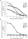
Fig 1. Neutralizing activities of phage library-derived IgGs against HCVpp infection.
The neutralization effects of the e2d066, e2d073, and e2d081 antibodies were assessed by infection assays using HCV pseudoparticles (HCVpp) harboring the E1 and E2 glycoproteins of (A) TH (genotype 1b) and (B) J6CF (genotype 2a). HCVpp, which include a luciferase-encoding construct, were mixed with immunoglobulin G (IgG) or phosphate buffered saline (PBS) for 30 minutes at room temperature; the mixtures then were used to inoculate naïve Huh-7.5.1 cells. Monoclonal anti-CD81 antibody (JS-81, BD Pharmingen) was used as positive control in this assay. At 72 hours after infection, the infected cells were harvested and lysed with Cell Culture Lysis Reagent (Promega). Luciferase activity was quantified using the Luciferase Assay System (Promega). Neutralizing activity was calculated and is presented as the % neutralization by comparison with the luciferase activities of the well inoculated with the HCVpp-PBS mixture. Assay were performed in triplicate and infection rate are expressed as mean ± SEM. Statistical significance of difference was analyzed using one-way ANOVA with a Williams’ test (*P < 0.025, **P < 0.005, ***P < 0.0005 vs PBS).
Fig 2
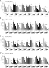
Fig 2. Epitope analysis with overlapping peptides.
A continuous series of TH E2 12-mer peptides (peptide Nos. 1 to 82), staggered by 3 amino acids each, were synthesized. Each peptide was modified to carry a biotinylation at the N-terminus and a glycine amide at the C-terminus. These peptides were used to coat a streptavidin-coated plate. (A) e2d066, (B) e2d073, (C) e2d081, or (D) MBL-HCV1 antibodies were applied to the peptide-coated plates. HRP-conjugated goat anti-human IgG antibody (Thermo Fisher Scientific) was used to detect the peptide-bound antibodies. The titers of bound antibodies were measured using a peroxidase assay kit (Type T; Sumitomo Bakelite Co., Ltd., Tokyo, Japan).
Fig 3
Fig 3. Antibody binding against naïve and denatured genotype 1b envelope E2 protein.
(A) Naïve and (B) denatured recombinant THE2-Fc proteins were used to coat Nunc-Immune plates (Thermo Fisher Scientific). Biotinylated anti-E2 antibodies were distributed to the prepared plates. Binding of biotinylated anti-E2 antibodies to the coated plates then was assessed using Streptavidin-HRP (GE Healthcare). The titers of antibodies were measured using a peroxidase assay kit for ELISA (Sumitomo Bakelite Co., Ltd.). Open-circle: e2d066 IgG, open-triangle: e2d073 IgG, open-square: e2d081 IgG, filled-circle: MBL-HCV1 (linear epitope control antibody), filled-triangle: AR3A (conformational epitope control antibody).
Fig 4
Fig 4. Competition assay using anti-E2 antibodies to compete against e2d066 IgG binding.
The TH E2-Fc recombinant E2 protein was used to coat Nunc-Immuno plates (Thermo Fisher Scientific). Biotinylated e2d066 IgG was mixed with each of the other anti-E2 antibodies, and the resulting mixtures were distributed to the prepared plates. Avidin-HRP (GE Healthcare) was used to detect the biotinylated IgGs. The titers of antibodies were measured using a peroxidase assay kit for ELISA (Sumitomo Bakelite Co., Ltd.). (A) Competition with linear epitope antibodies and AR3A. (B) Competition with the escape mutant-suppressive HC84-1 antibody. Open-circle: e2d066 IgG, open-triangle: e2d073 IgG, open-square: e2d081 IgG, filled-circle: MBL-HCV1, filled-triangle: AR3A, filled-rhombus: 8D10-3, filled-square: HC-84.1.
Fig 5
Fig 5. Neutralizing activity of anti-E2 IgG and scFv antibodies against HCVcc infection.
The neutralization effects of anti-E2 antibodies were assessed using an infection system with chimeric HCVcc, including (A) H77/JFH-1, (B) TH/JFH-1, (C) J6/JFH-1 and (D) S310/JFH-1. A total of 200 focus forming unit of these viruses were mixed with IgG or scFv at the designated concentrations and the mixtures were incubated at room temperature for 30 minutes. The antibody-HCVcc mixture was added to naïve Huh-7.5.1 cells (2 x 10⁴ cells/well, 48-well plate) at 100 μL/well and plates were incubated in a 5% CO2 incubator at 37°C for 3 hours. The antibody-HCVcc mixture then was removed and replaced with 500 μL of DMEM containing 10% FBS, and the cells were cultured in a 5% CO2 incubator at 37°C for a further 72 hours. The cells then were washed with PBS, and Passive Lysis Buffer (Promega) was added at 100 μL/well to generate a cell lysate. HCV core protein in the collected cell lysate was quantified using a Lumipulse Ortho HCV Ag (Ortho Clinical Diagnostics, Tokyo, Japan). The efficiency of neutralization was calculated and presented as the % neutralization normalized to the amount of HCV core protein in the HCVcc-PBS mixture-inoculated cell lysate. Assay were performed in triplicate and infection rate are expressed as mean ± SEM. Statistical significance of difference was analyzed using one-way ANOVA with a Williams’ test (*P < 0.025, **P < 0.005, ***P < 0.0005 vs PBS).

References

    1. Choo QL, Kuo G, Weiner AJ, Overby LR, Bradley DW, Houghton M. Isolation of a cDNA clone derived from a blood-borne non-A, non-B viral hepatitis genome. Science 1989;244:359–362. doi: 10.1126/science.2523562 - DOI - PubMed
    1. Hepatitis C fact sheet. World Health Organization. July 2021. Available from: https://www.who.int/news-room/fact-sheets/detail/hepatitis-c
    1. Wang CC, Krantz E, Klarquist J, Krows M, McBride L, Scott EP, et al.. Acute hepatitis C in a contemporary US cohort: modes of acquisition and factors influencing viral clearance. J Infect Dis 2007;196:1474–1482. doi: 10.1086/522608 - DOI - PubMed
    1. Grebely J, Page K, Sacks-Davis R, van der Loeff MS, Rice TM, Bruneau J, et al.. The effects of female sex, viral genotype, and IL28B genotype on spontaneous clearance of acute hepatitis C virus infection. Hepatology 2014;59:109–120. doi: 10.1002/hep.26639 - DOI - PMC - PubMed
    1. Osburn WO, Snider AE, Wells BL, Latanich R, Bailey JR, Thomas DL, et al.. Clearance of hepatitis C infection is associated with the early appearance of broad neutralizing antibody responses. Hepatology 2014;59:2140–2151. doi: 10.1002/hep.27013 - DOI - PMC - PubMed

Publication types