. 2022 Nov 21;35(11):2122-2132.
doi: 10.1021/acs.chemrestox.2c00231. Epub 2022 Sep 22.

Shared Clavulanate and Tazobactam Antigenic Determinants Activate T-Cells from Hypersensitive Patients

Affiliations

Shared Clavulanate and Tazobactam Antigenic Determinants Activate T-Cells from Hypersensitive Patients

Adriana Ariza et al. Chem Res Toxicol. .

Abstract

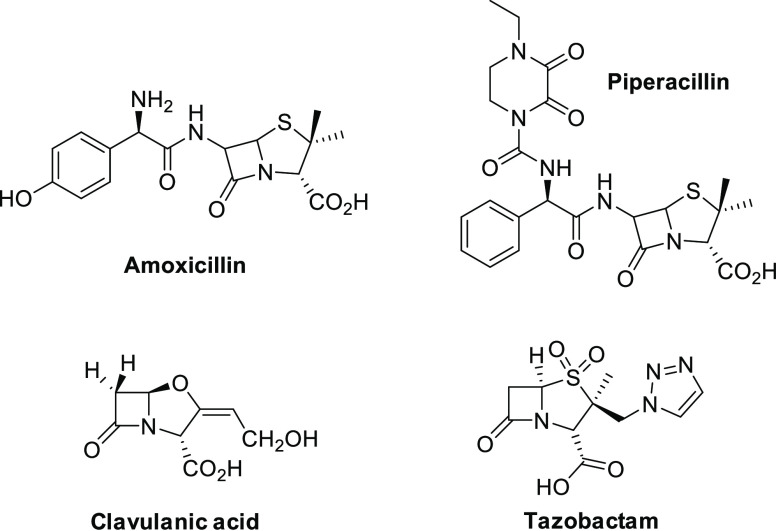
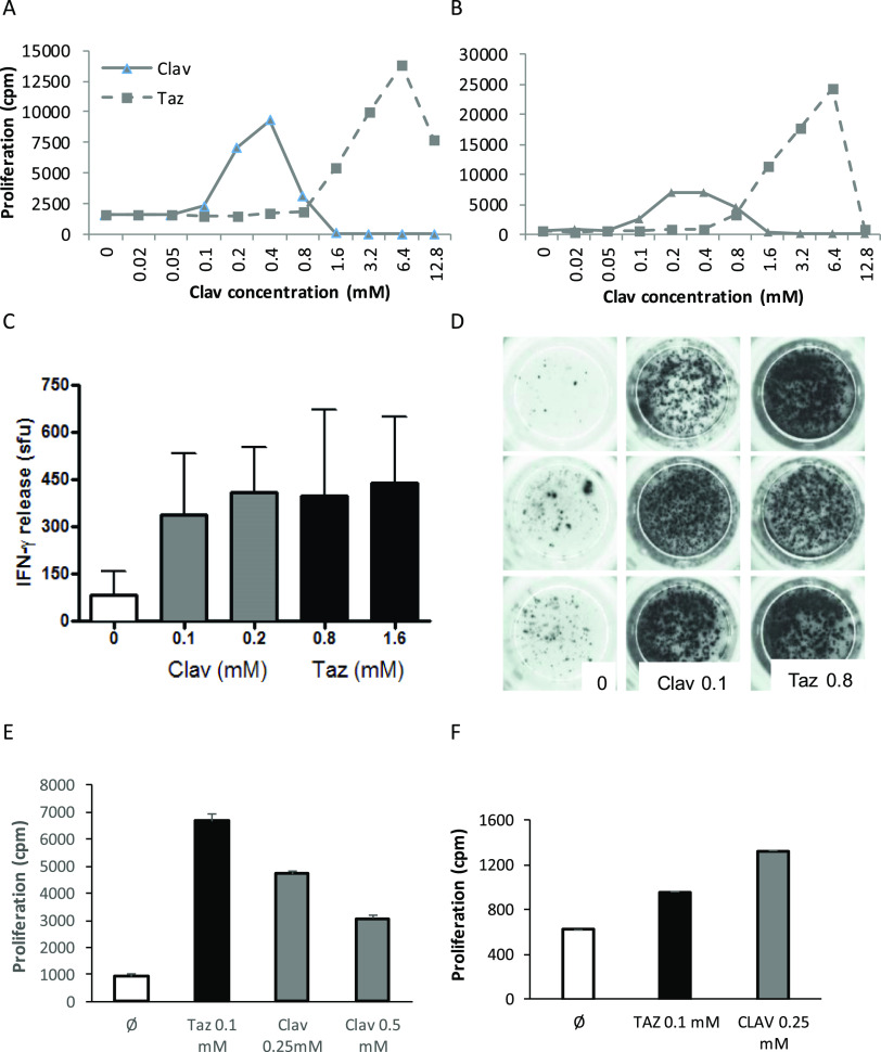
β-Lactamase inhibitors such as clavulanic acid and tazobactam were developed to overcome β-lactam antibiotic resistance. Hypersensitivity reactions to these drugs have not been studied in detail, and the antigenic determinants that activate T-cells have not been defined. The objectives of this study were to (i) characterize clavulanate- and tazobactam-responsive T-cells from hypersensitive patients, (ii) explore clavulanate and tazobactam T-cell crossreactivity, and (iii) define the antigenic determinants that contribute to T-cell reactivity. Antigen specificity, pathways of T-cell activation, and crossreactivity with clavulanate- and tazobactam-specific T-cell clones were assessed by proliferation and cytokine release assays. Antigenic determinants were analyzed by mass spectrometry-based proteomics methods. Clavulanate- and tazobactam-responsive CD4+ T-cell clones were stimulated to proliferate and secrete IFN-γ in an MHC class II-restricted and dose-dependent manner. T-cell activation with clavulanate- and tazobactam was dependent on antigen presenting cells because their fixation prevented the T-cell response. Strong crossreactivity was observed between clavulanate- and tazobactam-T-cells; however, neither drug activated β-lactam antibiotic-responsive T-cells. Mass spectrometric analysis revealed that both compounds form multiple antigenic determinants with lysine residues on proteins, including an overlapping aldehyde and hydrated aldehyde adduct with mass additions of 70 and 88 Da, respectively. Collectively, these data show that although clavulanate and tazobactam are structurally distinct, the antigenic determinants formed by both drugs overlap, which explains the observed T-cell cross-reactivity.


Conflict of interest statement

The authors declare no competing financial interest.

Figures

Figure 1
Figure 1
Chemical structure of B-lactam antibiotics and B-lactamase inhibitors.
Figure 2
Figure 2
T-cells from clavulanic acid or tazobactam hypersensitivity patients are stimulated by clavulanate or tazobactam. T-cell clones were generated from the blood of clavulanic acid or tazobactam hypersensitive patients. Clones were then cultured with autologous APCs in the presence of medium as a negative control and clavulanate (A–C) or tazobactam (D) for 2 days. Proliferation was measured by the addition of [3H]-thymidine. Results are expressed as the SI, calculated by dividing the mean counts per minute (cpm) of drug-stimulated cells by the mean cpm of nonstimulated cells. Clones were expanded for further assessment when SI > 1.5 was recorded.
Figure 3
Figure 3
Clavulanate- and tazobactam-responsive T-cell clones are activated with the alternative 6-lactamase inhibitor. (AB) Dose-dependent proliferation of clavulanate T-cell clones with clavulanate and tazobactam. Clones were cultured with autologous APCs in the presence of medium as a negative control and titrated concentrations of the B-lactamase inhibitors for 2 days before the addition of [3H]-thymidine. Results are expressed as the mean counts per minute (cpm). (C,D) Clavulanate T-cell clones were cultured with APCs and clavulanate or tazobactam and IFN-P secretion was measured using ELIspot. (E,F) Tazobactam T-cell clones were stimulated to proliferate in the presence of tazobactam and clavulanate. Proliferation was measured by the addition of [3H]-thymidine.
Figure 4
Figure 4
Crossreactivity of 6-lactam antibiotic-responsive T-cell clones with clavulanate or tazobactam. Proliferation assay showing that (A) amoxicillin- and (B) piperacillin-specific T-cell clones are not activated with either clavulanate or tazobactam. T-cell clones were cultured with APCs and either amoxicillin, clavulanate, tazobactam, or piperacillin for 2 days before analysis of proliferation by the addition of [3H]-thymidine. Results are expressed as mean counts per minute (cpm).
Figure 5
Figure 5
Tazobactam stimulates T-cell clones via an MHC-restricted and processing-dependent pathway. (A) T-cell clones were cocultured with drug and APC (+APC), glutaraldehyde-fixed APC (+Fix APC) or in the absence of APC (−APC) for 2 days before proliferation analysis by the addition of [3H]thymidine. (B) Proliferation assay to analyze tazobactam presentation to T-cell clones in the presence and absence of the MHC block. APC was incubated for 30 min at 37 °C with MHC class I and II blocking antibodies or isotype control before being cultured with drug and T-cell clones for 2 days. Proliferation was measured by the addition of [3H]thymidine. Results are expressed as counts per minute (cpm).
Figure 6
Figure 6
LC–MS/MS characterization of HSA adducts formed by tazobactam and clavulanate in vitro. Representative MS/MS spectrum of the albumin peptide 182LDELRDEGKASSAK195 modified at Lys190 with clavulanate (A). A similar adduct was also formed by tazobactam (B). The epitope map on HSA shows the lysine residues modified by tazobactam (at drug/protein molar ratio 100:1) or clavulanate (at drug/protein molar ratio 20:1); residues modified by both compounds are highlighted in pink and additional residues modified only by clavulanate are highlighted in green. Images are illustrated by PyMOL (The PyMOL Molecular Graphics System, Version 1.3 Schrödinger, LLC). The level of modification with clavulanate and tazobactam is concentration-dependent (D,E). (F) Summary of the HSA lysine residues targeted by clavulanate and tazobactam and the nature of the adducts formed.
Figure 7
Figure 7
MS/MS spectra of tazobactam-modified peptides. A representative MS/MS spectrum shows a HSA peptide K*QTALVELVK that was modified by tazobactam with a mass addition of 88 Da (A). A similar adduct was also detected when clavulanate was incubated with HSA (B). Albumin peptides modified by clavulanate or tazobactam were identified in vitro. Clavulanate and tazobactam were incubated with HSA for 24 h at a drug protein molar ratio of 20:1 and 200:1, respectively.
Scheme 1
Scheme 1. (A) Degradation of Covalently Bound Clavulanate to Lys19 and (B) Further hydrolysis Resulting in an Aldehyde/Hydrated Aldehyde Adduct

References

    1. Stover K. R.; Barber K. E.; Wagner J. L. Allergic reactions and cross-reactivity potential with beta-lactamase inhibitors. Pharmacy 2019, 7, 77.10.3390/pharmacy7030077. - DOI - PMC - PubMed
    1. Drawz S. M.; Bonomo R. A. Three decades of beta-lactamase inhibitors. Clin. Microbiol. Rev. 2010, 23, 160–201. 10.1128/CMR.00037-09. - DOI - PMC - PubMed
    1. Finlay J.; Miller L.; Poupard J. A. A review of the antimicrobial activity of clavulanate. J. Antimicrob. Chemother. 2003, 52, 18–23. 10.1093/jac/dkg286. - DOI - PubMed
    1. Gavin P. J.; Suseno M. T.; Thomson R. B. Jr.; Gaydos J. M.; Pierson C. L.; Halstead D. C.; Aslanzadeh J.; Brecher S.; Rotstein C.; Brossette S. E.; Peterson L. R. Clinical correlation of the CLSI susceptibility breakpoint for piperacillin- tazobactam against extended-spectrum-beta-lactamase-producing Escherichia coli and Klebsiella species. Antimicrob. Agents Chemother. 2006, 50, 2244–2247. 10.1128/AAC.00381-05. - DOI - PMC - PubMed
    1. Jones R. N.; Pfaller M. A.; Fuchs P. C.; Aldridge K.; Allen S. D.; Gerlach E. H. Piperacillin/tazobactam (YTR 830) combination. Comparative antimicrobial activity against 5889 recent aerobic clinical isolates and 60 Bacteroides fragilis group strains. Diagn. Microbiol. Infect. Dis. 1989, 12, 489–494. 10.1016/0732-8893(89)90083-7. - DOI - PubMed

Publication types