

Beyond Simpson's Rule: Accounting for Orientation and Ellipticity Assumptions
- PMID: 36137846
- PMCID: PMC9810537
- DOI: 10.1016/j.ultrasmedbio.2022.07.013
Beyond Simpson's Rule: Accounting for Orientation and Ellipticity Assumptions
Abstract
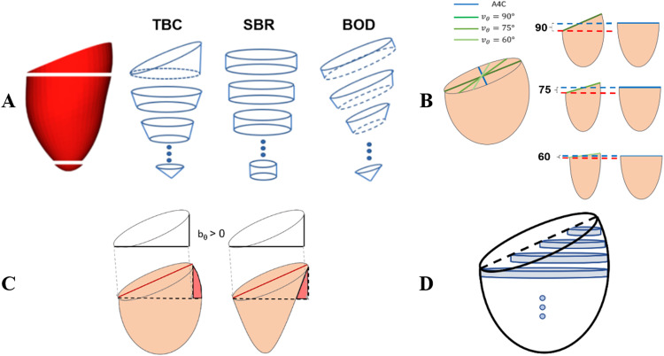
Simpson's biplane rule (SBR) is considered the gold standard method for left ventricle (LV) volume quantification from echocardiography but relies on a summation-of-disks approach that makes assumptions about LV orientation and cross-sectional shape. We aim to identify key limiting factors in SBR and to develop a new robust standard for volume quantification. Three methods for computing LV volume were studied: (i) SBR, (ii) addition of a truncated basal cone (TBC) to SBR and (iii) a novel method of basal-oriented disks (BODs). Three retrospective cohorts representative of the young, adult healthy and heart failure populations were used to study the impact of anatomical variations in volume computations. Results reveal how basal slanting can cause over- and underestimation of volume, with errors by SBR and TBC >10 mL for slanting angles >6°. Only the BOD method correctly accounted for basal slanting, reducing relative volume errors by SBR from -2.23 ± 2.21% to -0.70 ± 1.91% in the adult population and similar qualitative performance in the other two cohorts. In conclusion, the summation of basal oriented disks, a novel interpretation of SBR, is a more accurate and precise method for estimating LV volume.
Keywords: Apical chamber views; Left ventricle volumes; Modified Simpson's biplane rule; Two-dimensional echocardiography.
Copyright © 2022 The Authors. Published by Elsevier Inc. All rights reserved.
Conflict of interest statement
Conflict of interest disclosure W.J.C.K. is a PhD student funded at a 50% level by Ultromics Ltd. P. Leeson is the Academic Founder and Non-Executive Director of Ultromics. A.B. and D.M. are Ultromics employees. P. Lamata is a member of the Ultromics Advisory Board. Ultromics is not seeking to protect the intellectual property of the BOD formulation, hoping that all vendors quickly adopt it.
Figures


References
-
- Baicu CF, Zile MR, Aurigemma GP, Gaasch WH. Left ventricular systolic performance, function, and contractility in patients with diastolic heart failure. Circulation. 2005;111:2306–2312. - PubMed
-
- Bellenger N. Comparison of left ventricular ejection fraction and volumes in heart failure by echocardiography, radionuclide ventriculography and cardiovascular magnetic resonance. Are they interchangeable? Eur Heart J. 2000;21:1387–1396. - PubMed
-
- Bonhorst D, Guerreiro S, Fonseca C, Cardim N, Macedo F, Adragão P. Real-life data on heart failure before and after implantation of resynchronization and/or defibrillation devices— The Síncrone study. Rev Port Cardiol (Engl Ed) 2019;38:33–41. - PubMed
-
- Chan K, Mahmod M, Zacur E, Rigolli M, Francis J, Ariga R, Raman B, Dass S, Karamitsos T, Neubauer S, Myerson S, Lamata P. Reappraising remodelling pattern of left ventricle in aortic stenosis: Axis orientation as a unique signature of positive remodelling. Heart. 2019;105(Suppl 6):A18.
Publication types
MeSH terms
Associated data
Grants and funding
LinkOut - more resources
Full Text Sources
