. 2022 Nov;127(10):1893-1905.
doi: 10.1038/s41416-022-01966-5. Epub 2022 Sep 22.

FAK promotes stromal PD-L2 expression associated with poor survival in pancreatic cancer

Affiliations

FAK promotes stromal PD-L2 expression associated with poor survival in pancreatic cancer

Catherine Davidson et al. Br J Cancer. 2022 Nov.

Abstract

Background: Pancreatic Cancer is one of the most lethal cancers, with less than 8% of patients surviving 5 years following diagnosis. The last 40 years have seen only small incremental improvements in treatment options, highlighting the continued need to better define the cellular and molecular pathways contributing to therapy response and patient prognosis.

Methods: We combined CRISPR, shRNA and flow cytometry with mechanistic experiments using a KrasG12Dp53R172H mouse model of pancreatic cancer and analysis of publicly available human PDAC transcriptomic datasets.

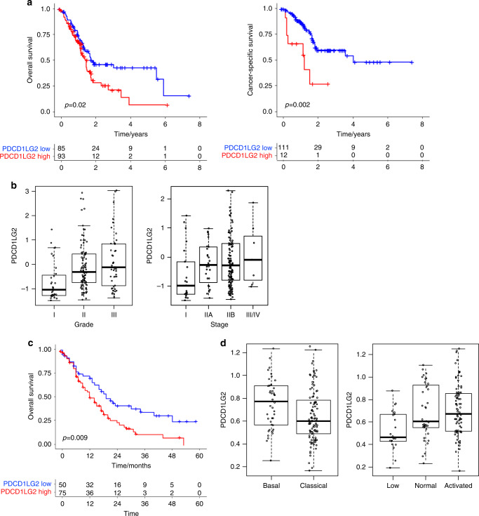
Results: Here, we identify that expression of the immune checkpoint, Programmed Death Ligand 2 (PD-L2), is associated with poor prognosis, tumour grade, clinical stage and molecular subtype in patients with Pancreatic Ductal Adenocarcinoma (PDAC). We further show that PD-L2 is predominantly expressed in the stroma and, using an orthotopic murine model of PDAC, identify cancer cell-intrinsic Focal Adhesion Kinase (FAK) signalling as a regulator of PD-L2 stromal expression. Mechanistically, we find that FAK regulates interleukin-6, which can act in concert with interleukin-4 secreted by CD4 T-cells to drive elevated expression of PD-L2 on tumour-associated macrophages, dendritic cells and endothelial cells.

Conclusions: These findings identify further complex heterocellular signalling networks contributing to FAK-mediated immune suppression in pancreatic cancer.


Conflict of interest statement

The authors declare no competing interests.

Figures

Fig. 1
Fig. 1. FAK promotes murine PDAC growth associated with increased stromal expression of PD-L2.
a Representative western blot of Panc47 FAK-wt and FAK−/− whole cell lysates probed with anti-FAK and anti-GAPDH antibodies. b Average weight of Panc47 FAK-wt and FAK−/− tumours 2 and 3 weeks post-implantation of 0.5 × 106 cells into the pancreas of C57BL/6 mice. n = 8 tumours per group. c Kaplan–Meier survival plot of C57BL/6 mice implanted with Panc47 FAK-wt and FAK−/− tumours. n = 8 mice per group. Log-rank (Mantel-Cox) test, p = 0.0005; Gehan-Breslow-Wilcoxon test, p = 0.0015. d Nanostring gene expression analysis of RNA isolated from Panc47 FAK-wt and FAK−/− tumours. n = 3 tumours per group. e t-sne map of flow cytometry data from Panc47 FAK-wt and Panc47 FAK−/− tumours. Data generated from all live cells in a representative tumour. f Flow cytometry quantification of PD-L2 expression in Panc47 FAK-wt and FAK−/− tumours. n = 9 tumours per group. Data represented as mean ± s.e.m. Two-tailed unpaired t-test, ****p ≤ 0.0001, ***p ≤ 0.001, **p ≤ 0.01, *p ≤ 0.05.
Fig. 2
Fig. 2. PD-L2 expression is associated with tumour grade, molecular subtype and poor patient survival in PDAC.
a Kaplan–Meier plot of overall survival (left) and cancer-specific survival (right) in PDAC patients with low and high expression of PDCD1LG2. Source data: TCGA. b Left—PDCD1LG2 expression in grade I–III PDAC tumours. Source data: TCGA. Grade I vs III, p = 0.0001. right—PDCD1LG2 expression in stage I–IV PDAC tumours. Source data: TCGA. Grade I vs all other stages, p = 0.004. c Kaplan–Meier plot of survival probability in PDAC patients with low and high expression of PDCD1LG2. Source data: Moffit et al. d Left—PDCD1LG2 expression in Classical vs Basal PDAC tumour subtypes. Source data: Moffit et al. p = 0.01. right—PDCD1LG2 expression in low, normal and activated stromal PDAC tumour subtypes. Source data: Moffit et al. low vs activated p = 0.0005.
Fig. 3
Fig. 3. PD-L2 is predominantly expressed in in human PDAC stroma.
a PDCD1LG2 expression in LCM separated RNASeq analysis of 66 pairs of Epithelium and Stroma and 15 bulk human PDAC [32]. be Co-expression of PDCD1LG2 and genes associated with immune cell subsets in TCGA [28] and Moffitt et al. [29] bulk RNAseq datasets from primary human PDAC.
Fig. 4
Fig. 4. FAK-dependent expression of stromal PD-L2 requires CD4+ T cells.
a Flow cytometry quantification of PD-L2 expression on bone-marrow derived macrophages cultured in normal growth media (M), FAK-wt conditioned media (CM) or M / CM supplemented with IL4. n = 4 per condition. Ordinary one-way ANOVA with Tukey’s multiple comparison. b Analysis of the correlation between CD4 and PDCD1LG2 gene expression in human PDAC RNAseq data (TCGA, 184 samples). c Flow cytometry quantification of CD45+CD3+CD4+IL4+ cells as a percentage of CD45+ cells in Panc47 FAK-wt and FAK−/− tumours. n = 6 per condition. d Left—Flow cytometry quantification of the median fluorescence intensity of IL4 expression in CD45+CD3+CD4+IL4+ cells from Panc47 FAK-wt and FAK−/− tumours. Data represented as a fold-change relative to FAK-wt. n = 6 per condition. Right—representative histogram showing IL4 expression compared to FMO control. e Fold-change in average weight of Panc47 FAK-wt and FAK−/− tumours following treatment with either isotype control or anti-CD4 antibodies. n = 3, one-way ANOVA Fisher’s LSD. f Flow cytometry quantification of PD-L2 expression in Panc47 FAK-wt tumours following treatment with either isotype control antibody or anti-CD4 T-cell depleting antibody. n = 6, two-tailed unpaired t-test. All data represented as mean ± s.e.m. ****p ≤ 0.0001, ***p ≤ 0.001, **p ≤ 0.01, *p ≤ 0.05.
Fig. 5
Fig. 5. FAK-dependent expression of IL6 amplifies IL4-dependent PD-L2 expression.
a Quantitative analysis of chemokines / cytokines present in media conditioned by either Panc47 FAK-wt or Panc47 FAK−/− cells for 48 h. b Flow cytometry quantification of PD-L2 expression on bone-marrow derived macrophages cultured in normal growth media (M), M + IL6, M + IL4 and M + IL6 + IL4. Data represented as fold-change in median fluorescence intensity relative of M alone. n = 3 per condition, ordinary one-way ANOVA with Tukey’s multiple comparison. c ELISA quantification of IL6 in media conditioned by either Panc47 FAK-wt or Panc47 FAK−/− cells untreated or stimulated with IL17. n = 3, two-tailed unpaired t-test. d ELISA quantification of IL6 in media conditioned by either Panc47 FAK-wt (n = 7), FAK−/− (n = 7), FAK-wt control shRNA (n = 7), FAK-wt IL6 shRNA1 (n = 6) and FAK-wt IL6 shRNA2 cells (n = 6). One-way ANOVA with Dunnett’s multiple comparison. e Fold-change in the average weight of Panc47 FAK-wt (n = 14), FAK−/− (n = 11), FAK-wt control shRNA (n = 4), FAK-wt IL6 shRNA1 (n = 6) and FAK-wt IL6 shRNA2 (n = 6) tumours 2 weeks post-implantation of 0.5 × 106 cells into the pancreas of C57BL/6 mice. Ordinary one-way ANOVA with Tukey’s multiple comparison. f Flow cytometry quantification of PD-L2 expression in Panc47 FAK-wt CTL shRNA (n = 7), FAK-wt IL6 shRNA1 (n = 3) and FAK-wt IL6 shRNA2 (n = 3) tumours. Ordinary one-way ANOVA with Tukey’s multiple comparison. All data represented as mean ± s.e.m. ****p ≤ 0.0001, ***p ≤ 0.001, **p ≤ 0.01, *p ≤ 0.05.
Fig. 6
Fig. 6. Model.
Graphical summary of proposed mechanism through which FAK-IL6 signaling amplifies PD-L2 expression in the PDAC stroma. Image created using BioRender.

References

    1. Siegel RL, Miller KD, Jemal A. Cancer statistics, 2018. CA Cancer J Clin. 2018;68:7–30. doi: 10.3322/caac.21442. - DOI - PubMed
    1. Ferlay J, Partensky C, Bray F. More deaths from pancreatic cancer than breast cancer in the EU by 2017. Acta Oncol. 2016;55:1158–60. doi: 10.1080/0284186X.2016.1197419. - DOI - PubMed
    1. Rahib L, Smith BD, Aizenberg R, Rosenzweig AB, Fleshman JM, Matrisian LM. Projecting cancer incidence and deaths to 2030: the unexpected burden of thyroid, liver, and pancreas cancers in the United States. Cancer Res. 2014;74:2913–21. doi: 10.1158/0008-5472.CAN-14-0155. - DOI - PubMed
    1. Balachandran VP, Beatty GL, Dougan SK. Broadening the impact of immunotherapy to pancreatic cancer: challenges and opportunities. Gastroenterology. 2019;156:2056–72. doi: 10.1053/j.gastro.2018.12.038. - DOI - PMC - PubMed
    1. Neoptolemos JP, Kleeff J, Michl P, Costello E, Greenhalf W, Palmer DH. Therapeutic developments in pancreatic cancer: current and future perspectives. Nat Rev Gastroenterol Hepatol. 2018;15:333–48. doi: 10.1038/s41575-018-0005-x. - DOI - PubMed

Publication types

MeSH terms

Substances