

Radiological Characteristics of Low-Grade Lytic Spondylolisthesis: Similarity to Dysplastic Spondylolisthesis
- PMID: 36138575
- PMCID: PMC10151640
- DOI: 10.31616/asj.2022.0033
Radiological Characteristics of Low-Grade Lytic Spondylolisthesis: Similarity to Dysplastic Spondylolisthesis
Abstract
Study design: Retrospective case-control study.
Purpose: This study aimed to analyze the etiology of low-grade lytic spondylolisthesis based on the radiologic features of the vertebra.
Overview of literature: According to the Marchetti-Bartolozzi classification scheme, high-grade lytic spondylolisthesis (Meyerding grade 3-5) is classified as dysplastic. However, determination of the etiology for low-grade lytic spondylolisthesis as developmental or traumatic remains controversial.
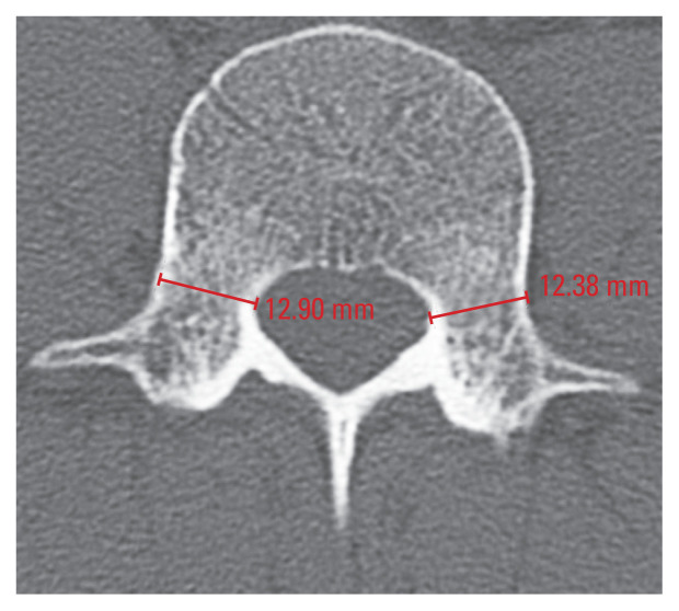
Methods: Patients admitted and treated for one-level (L4/5 or L5/S1) low-grade spondylolisthesis were included in the study. A total of 135 patients were divided into the degenerative or lytic spondylolisthesis groups according to their condition (81 patients [degenerative group] vs. 54 patients [lytic group]). To assess the level of similarity in the radiological findings between low-grade lytic spondylolisthesis and dysplastic spondylolisthesis, the pedicle diameters and vertebral heights of the L4 and L5 vertebrae were measured on computed tomography images. Measurements were then converted to each vertebra's ratio to reduce confounding factors among individuals.
Results: The affected vertebra had a smaller sagittal pedicle diameter/transverse pedicle diameter ratio in the low-grade lytic spondylolisthesis group compared to the degenerative group, and the posterior vertebral height/anterior vertebral height ratio of L5 was smaller in the L5/S1 lytic spondylolisthesis group compared to the degenerative spondylolisthesis group.
Conclusions: Low-grade lytic spondylolisthesis and dysplastic spondylolisthesis demonstrated similar radiological findings. Hence, surgeons should be attentive to the morphology of the vertebral body and posterior column during preoperative planning for the treatment of low-grade lytic spondylolisthesis.
Keywords: Dysplasia; Lumbar vertebrae; Spondylolisthesis; Vertebral body; Vertebral pedicles.
Conflict of interest statement
No potential conflict of interest relevant to this article was reported.
Figures


Similar articles
-
A radiological parametric comparison of low-grade lytic spondylolisthesis to degenerative spondylolisthesis - A retrospective approach to establish its dysplastic origin.J Craniovertebr Junction Spine. 2024 Jan-Mar;15(1):30-36. doi: 10.4103/jcvjs.jcvjs_136_23. Epub 2024 Mar 13. J Craniovertebr Junction Spine. 2024. PMID: 38644923 Free PMC article.
-
Anatomical parameters of fifth lumbar vertebra in L5-S1 spondylolytic spondylolisthesis from a surgical point of view.Eur Spine J. 2014 Sep;23(9):1896-902. doi: 10.1007/s00586-013-3111-z. Epub 2013 Nov 26. Eur Spine J. 2014. PMID: 24275826
-
What is the difference in pedicle morphology of the fifth lumbar vertebra between isthmic and degenerative L5-S1 spondylolisthesis? An anatomic study of 328 patients via multi-slice spiral computed tomography.Eur Spine J. 2021 Aug;30(8):2301-2310. doi: 10.1007/s00586-021-06884-3. Epub 2021 May 28. Eur Spine J. 2021. PMID: 34050393
-
Management of High-Grade Dysplastic Spondylolisthesis.Neurosurg Clin N Am. 2023 Oct;34(4):567-572. doi: 10.1016/j.nec.2023.06.003. Epub 2023 Jul 23. Neurosurg Clin N Am. 2023. PMID: 37718103 Review.
-
Degenerative lumbar spondylolisthesis: cohort of 670 patients, and proposal of a new classification.Orthop Traumatol Surg Res. 2014 Oct;100(6 Suppl):S311-5. doi: 10.1016/j.otsr.2014.07.006. Epub 2014 Sep 5. Orthop Traumatol Surg Res. 2014. PMID: 25201282 Review.
Cited by
-
A radiological parametric comparison of low-grade lytic spondylolisthesis to degenerative spondylolisthesis - A retrospective approach to establish its dysplastic origin.J Craniovertebr Junction Spine. 2024 Jan-Mar;15(1):30-36. doi: 10.4103/jcvjs.jcvjs_136_23. Epub 2024 Mar 13. J Craniovertebr Junction Spine. 2024. PMID: 38644923 Free PMC article.
References
-
- Niggemann P, Kuchta J, Beyer HK, Grosskurth D, Schulze T, Delank KS. Spondylolysis and spondylolisthesis: prevalence of different forms of instability and clinical implications. Spine (Phila Pa 1976) 2011;36:E1463–8. - PubMed
-
- Hammerberg KW. New concepts on the pathogenesis and classification of spondylolisthesis. Spine (Phila Pa 1976) 2005;30(6 Suppl):S4–11. - PubMed
-
- Wiltse LL. The etiology of spondylolisthesis. J Bone Joint Surg Am. 1962;44-A:539–60. - PubMed
-
- Ikata T, Miyake R, Katoh S, Morita T, Murase M. Pathogenesis of sports-related spondylolisthesis in adolescents: radiographic and magnetic resonance imaging study. Am J Sports Med. 1996;24:94–8. - PubMed
LinkOut - more resources
Full Text Sources
Research Materials
