Review
. 2022 Sep 14;11(9):1351.
doi: 10.3390/biology11091351.

Molecular Mechanism of Blood Pressure Regulation through the Atrial Natriuretic Peptide

Affiliations
Review

Molecular Mechanism of Blood Pressure Regulation through the Atrial Natriuretic Peptide

Takeshi Tokudome et al. Biology (Basel). .

Abstract

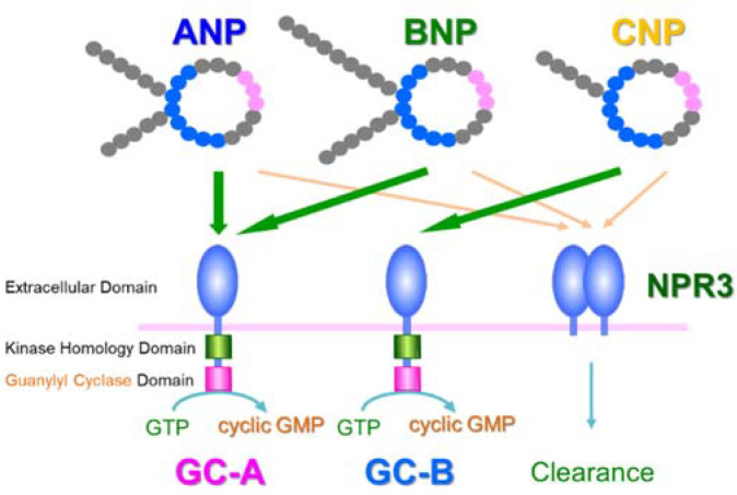
Natriuretic peptides, including atrial natriuretic peptide (ANP), brain natriuretic peptide (BNP), and C-type natriuretic peptide (CNP), have cardioprotective effects and regulate blood pressure in mammals. ANP and BNP are hormones secreted from the heart into the bloodstream in response to increased preload and afterload. Both hormones act through natriuretic peptide receptor 1 (NPR1). In contrast, CNP acts through natriuretic peptide receptor 2 (NPR2) and was found to be produced by the vascular endothelium, chondrocytes, and cardiac fibroblasts. Based on its relatively low plasma concentration compared with ANP and BNP, CNP is thought to function as both an autocrine and a paracrine factor in the vasculature, bone, and heart. The cytoplasmic domains of both NPR1 and NPR2 display a guanylate cyclase activity that catalyzes the formation of cyclic GMP. NPR3 lacks this guanylate cyclase activity and is reportedly coupled to Gi-dependent signaling. Recently, we reported that the continuous infusion of the peptide osteocrin, an endogenous ligand of NPR3 secreted by bone and muscle cells, lowered blood pressure in wild-type mice, suggesting that endogenous natriuretic peptides play major roles in the regulation of blood pressure. Neprilysin is a neutral endopeptidase that degrades several vasoactive peptides, including natriuretic peptides. The increased worldwide clinical use of the angiotensin receptor-neprilysin inhibitor for the treatment of chronic heart failure has brought renewed attention to the physiological effects of natriuretic peptides. In this review, we provide an overview of the discovery of ANP and its translational research. We also highlight our recent findings on the blood pressure regulatory effects of ANP, focusing on its molecular mechanisms.

Keywords: atrial natriuretic peptide; blood pressure; heart failure; molecular mechanism; translational research.


Conflict of interest statement

The authors declare no conflict of interest.

Figures

Figure 1
Figure 1
Natriuretic peptides and their receptors. ANP—atrial natriuretic peptide; BNP—brain natriuretic peptide; CNP—C-type natriuretic peptide; GC-A—guanylyl cyclase-A; GC-B—guanylyl cyclase-B; NPR3—natriuretic peptide receptor 3.
Figure 2
Figure 2
GC-A is abundantly expressed in rat small arteries and arterioles. Localization of GC-A in the aorta (A), mesenterium (B), and skeletal muscle (C) was examined by immunohistochemistry in 4-week-old male Sprague-Dawley rats. The region within the red dotted box in the upper part of each panel is magnified in the bottom part of each panel. This figure is a reconstruction of the figure in our original article [37].
Figure 3
Figure 3
GC-A in vascular endothelial cells contributes to hypotension induced by ANP infusion. Intravenous infusion of ANP (10 μg/kg/min) significantly lowered systolic blood pressure (SBP) compared with the administration of vehicle (saline) in wild-type mice (A), GC-A floxed mice (GC-Aflox/flox), (B), and smooth-muscle-cell-specific GC-A knockout mice (SMC-GC-A-KO), (C), but not in endothelial-cell-specific GC-A knockout mice (EC-GC-A-KO), (D). * p < 0.05 vs. vehicle. This figure is a reconstruction of the figure from our original article [37].
Figure 4
Figure 4
ANP-induced hypotension is independent of nitric oxide. (A) The measurement of nitric oxide in human umbilical vein endothelial cells (HUVECs). HUVECs were loaded with DAF-2 DA (a fluorescent nitric oxide probe) and then treated with vehicle (PBS), ANP, VEGF, or VEGF + L-NAME. Nitric oxide production was measured as cellular fluorescence (arbitrary units). Values are expressed relative to the fluorescence intensity before vehicle treatment. (B) Intravenous infusion of ANP (10 μg/kg/min) significantly lowered systolic blood pressure (SBP) compared with the administration of vehicle (saline) in Nos3-knockout mice (Nos3-KO). * p < 0.05 vs. vehicle. This figure is a reconstruction of the figure from our original article [37].
Figure 5
Figure 5
ANP causes hyperpolarization of cultured vascular endothelial cells in a K+ channel-dependent manner via the GC-A–PKG1 signaling pathway. (A) ANP significantly decreased the membrane potential of human umbilical vein endothelial cells (HUVECs) compared with that in the human coronary artery smooth muscle cells (HCASMCs). Changes in the membrane potential were evaluated by monitoring the cellular fluorescence intensity. Values are expressed relative to the intensity at time 0. * p < 0.05 vs. HCASMCs. (B) ANP had no effect on the intracellular calcium concentration of HUVECs. Fura-2-AM-loaded HUVECs were stimulated with or without ANP, and cellular fluorescence was monitored for 120 s. GSK101 (TRPV4 agonist) was used as the positive control. * p < 0.05 vs. without Ca2+. (C) Intravenous infusion of ANP (10 μg/kg/min) significantly lowered systolic blood pressure (SBP) compared with vehicle (saline) administration in intermediate/small-conductance KCa (Kcnn4) knockout mice. * p < 0.05 vs. vehicle. (D) ANP caused hyperpolarization of cultured HUVECs in a K+ channel-dependent manner via the NPR1–PKG1 signaling pathway. HUVECs were treated with or without ANP (100 nM) or 8-Br-PET-cGMP (PKG1 activator; 200 μM) in the presence or absence of a non-selective K+ channel blocker TEA (tetraethylammonium; 10 mM), PKG inhibitor (Rp-8-Br-PET-cGMPS; 200 μM), or PKG1 siRNA for 300 s. * p < 0.05. p < 0.01 vs. vehicle. This figure is a reconstruction of the figure from our original article [37].
Figure 6
Figure 6
RGS2 plays a key role in the hypotensive effect of ANP infusion. (A) ANP increased Rgs2 mRNA expression in human umbilical vein endothelial cells (HUVECs) but not in human coronary artery smooth muscle cells (HCASMCs). HUVECs and HCASMCs were treated with vehicle (PBS) or ANP for 60 min. mRNA levels were normalized to those of GAPDH. Values are expressed relative to vehicle-treated cells. * p < 0.05. (B) ANP increased Rgs2 mRNA expression in CD31+ cells but not in CD31 cells. After 48 h of continuous administration of vehicle (saline) or ANP (0.2 μg/kg/min), the Rgs2 mRNA levels were evaluated in CD31+ cells and CD31 cells. (C) RGS2 plays a key role in ANP-mediated hyperpolarization in HUVECs. Changes in the membrane potential in HUVECs transfected with control siRNA or Rgs2 siRNA (siRgs2) were evaluated. DiBAC4(3)-loaded HUVECs were treated with ANP. Values are expressed relative to the intensity of the control siRNA-transfected cells at time 0. * p < 0.05 vs. siRgs2. (D) ANP infusion failed to decrease systolic blood pressure (SBP) in RGS2 knockout (Rgs2-KO) mice. Intravenous infusion of ANP (10 μg/kg/min) did not significantly lower SBP compared with the vehicle infusion. This figure is a reconstruction of the figure in our original article [37].
Figure 7
Figure 7
Proposed mechanism underlying the acute blood pressure decrease induced by the administration of ANP. The hypotensive effect of ANP is independent of nitric oxide and can be caused, at least in part, by endothelial GC-A-mediated, RGS2-dependent endothelial hyperpolarization. The exact mechanism of the production of the RGS2-induced endothelium-derived hyperpolarizing factor and subsequent hyperpolarization of smooth muscle cells requires further investigation. This figure is a reconstruction of the figure in our original article [37].
Figure 8
Figure 8
Endothelium-specific overexpression of GC-A results in hypotension and vasodilation in mice. (A) The plasma cyclic GMP level was higher in endothelial-cell-specific GC-A-overexpressing (Tg) mice than in wild-type (WT) mice. (B) The mean blood pressure, as determined by radiotelemetry, was significantly lower in the Tg mice than in the WT mice. (C) The heart weight to body weight ratio was significantly smaller in the Tg mice than in the WT mice. (D) Hemodynamic analysis revealed that the left ventricular (LV) maximum pressure and arterial elastance were lower in Tg mice than in the WT mice, but the cardiac output was comparable between the two types of mice. (E) Angiographic images from a WT mouse and a Tg mouse. Yellow arrows indicate a vessel that is dilated in the Tg relative to the WT. * p < 0.05. This figure is a reconstruction of the figure in our original article [37].

References

    1. Potter L.R., Abbey-Hosch S., Dickey D.M. Natriuretic peptides, their receptors, and cyclic guanosine monophosphate-dependent signaling functions. Endocr. Rev. 2006;27:47–72. doi: 10.1210/er.2005-0014. - DOI - PubMed
    1. Kishimoto I., Tokudome T., Nakao K., Kangawa K. Natriuretic peptide system: An overview of studies using genetically engineered animal models. FEBS J. 2011;278:1830–1841. doi: 10.1111/j.1742-4658.2011.08116.x. - DOI - PubMed
    1. Kuhn M. Molecular physiology of membrane guanylyl cyclase receptor. Physiol. Rev. 2016;96:751–804. doi: 10.1152/physrev.00022.2015. - DOI - PubMed
    1. Nakagawa Y., Nishikimi T., Kuwahara K. Atrial and brain natriuretic peptides: Hormones secreted from the heart. Peptides. 2019;111:18–25. doi: 10.1016/j.peptides.2018.05.012. - DOI - PubMed
    1. Kato J. Natriuretic peptides and neprilysin inhibition in hypertension and hypertensive organ damage. Peptides. 2020;132:170352. doi: 10.1016/j.peptides.2020.170352. - DOI - PubMed

LinkOut - more resources