. 2022 Aug 24;12(9):1169.
doi: 10.3390/biom12091169.

Renin-a in the Subfornical Organ Plays a Critical Role in the Maintenance of Salt-Sensitive Hypertension

Affiliations

Renin-a in the Subfornical Organ Plays a Critical Role in the Maintenance of Salt-Sensitive Hypertension

Silvana G Cooper et al. Biomolecules. .

Abstract

The brain renin-angiotensin system plays important roles in blood pressure and cardiovascular regulation. There are two isoforms of prorenin in the brain: the classic secreted form (prorenin/sREN) encoded by renin-a, and an intracellular form (icREN) encoded by renin-b. Emerging evidence indicates the importance of renin-b in cardiovascular and metabolic regulation. However, the role of endogenous brain prorenin in the development of salt-sensitive hypertension remains undefined. In this study, we test the hypothesis that renin-a produced locally in the brain contributes to the pathogenesis of hypertension. Using RNAscope, we report for the first time that renin mRNA is expressed in several regions of the brain, including the subfornical organ (SFO), the paraventricular nucleus of the hypothalamus (PVN), and the brainstem, where it is found in glutamatergic, GABAergic, cholinergic, and tyrosine hydroxylase-positive neurons. Notably, we found that renin mRNA was significantly elevated in the SFO and PVN in a mouse model of DOCA-salt-induced hypertension. To examine the functional importance of renin-a in the SFO, we selectively ablated renin-a in the SFO in renin-a-floxed mice using a Cre-lox strategy. Importantly, renin-a ablation in the SFO attenuated the maintenance of DOCA-salt-induced hypertension and improved autonomic function without affecting fluid or sodium intake. Molecularly, ablation of renin-a prevented the DOCA-salt-induced elevation in NADPH oxidase 2 (NOX2) in the SFO without affecting NOX4 or angiotensin II type 1 and 2 receptors. Collectively, our findings demonstrate that endogenous renin-a within the SFO is important for the pathogenesis of salt-sensitive hypertension.

Keywords: NAD(P)H oxidase; angiotensin receptor; autonomic control; renin-angiotensin system; salt-sensitive hypertension.


Conflict of interest statement

The authors declare that there are no competing interest associated with the manuscript.

Figures

Figure 1
Figure 1
Renin mRNA is upregulated in the SFO and PVN of DOCA-salt–treated mice. (AF) Total renin levels were detected in the subfornical organ (SFO), paraventricular nucleus of hypothalamus (PVN), arcuate nucleus (ARC), rostral ventrolateral medulla (RVLM), nucleus tractus solitarius (NTS), and area postrema (AP) using ddPCR. * p < 0.05 versus sham treatment (Student’s t test); n = 7–9 mice/group.
Figure 2
Figure 2
Cell type expression of renin in the SFO. Renin mRNA and mRNAs for nucleus-specific GABAergic and glutamatergic neuron markers were detected in the mouse brain (n = 4 mice) using RNAScope in situ hybridization. Cells were immunolabeled for astrocytes (GFAP) and microglia (Iba1) markers. (A) Representative confocal images of renin mRNA in the SFO and (B) scrambled renin probe (negative control). Magenta, renin; yellow, glutamatergic neurons (vGluT2); cyan, GABAergic neurons (VIAAT); green, astrocyte marker (GFAP) or microglia marker (Iba1). Tissues were counterstained with DAPI (blue). (C) Summary data showing the cellular distribution of renin mRNA in GABAergic and/or glutamatergic neurons. Abbreviations: SFO, subfornical organ; vGluT2, vesicular glutamate 2; VIAAT, vesicular inhibitory amino acid transporter; GFAP, glial fibrillary acidic protein; Iba1, ionized calcium binding adaptor molecule 1.
Figure 3
Figure 3
Characterization of SFO-targeted renin-a–KO mice. (A) Schematic illustrating Cre-loxP recombination in renin-a–floxed mice. (B) Schematic brain atlas and coordinates showing injection sites for AAV2-Cre-eGFP or AAV2-eGFP into the SFO and a representative image of eGFP expression in the SFO.
Figure 4
Figure 4
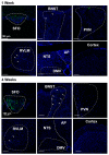
SFO-targeted AAV2-eGFP expression. Images of the SFO, cortex, BNST, PVN, RVLM, DMV, and NTS from mice that received an SFO injection of AAV2-eGFP 1 and 4 weeks after injections. eGFP expression (green) was detected in the cell bodies of SFO, but not in the bed of nucleus stria terminalis (BNST), paraventricular nucleus of the hypothalamus (PVN), rostral ventrolateral medulla (RVLM), nucleus tractus solitarius (NTS), dorsal motor nucleus of the vagus (DMV), area postrema (AP), or cortex. White arrows indicate visible projections (green dots, eGFP) to the BNST and RVLM. No obvious eGFP was observed in other brain regions examined.
Figure 5
Figure 5
Renin-a deletion in the SFO attenuates DOCA-salt–induced hypertension. Renin-a–floxed mice were injected in the SFO with either AAV2-eGFP or AAV2-Cre-eGFP. BP and HR were monitored by telemetry for 3 days before and 21 days after DOCA-salt treatment. (A) Experimental protocol for telemetric probe implantation, virus delivery, and DOCA-salt treatment. (B) Fluid intake of mice before and during 21-day DOCA-salt treatment (n = 8–10 mice/group). # p < 0.05, ## p < 0.01 versus AAV2-eGFP (two-way ANOVA with mixed-effects model, Fisher’s LSD test). (C) Continuous mean arterial pressure (MAP) recordings before and during 21-day DOCA-salt treatment (n = 15–18 mice/group). ## p < 0.01 versus AAV2-eGFP (two-way ANOVA with mixed-effects model, Fisher’s LSD test). (D) Endpoint MAP at 21 days DOCA-salt treatment (n = 7–15 mice/group). ** p < 0.01, **** p < 0.0001 versus baseline AAV2-eGFP; #### p < 0.0001 versus AAV2-eGFP DOCA-salt (one-way ANOVA with Fisher’s LSD test). (E) Continuous HR recording before and during 21-day DOCA-salt treatment (n = 11–20 mice/group). (F) Endpoint HR at 21 days DOCA-salt treatment (n = 11–20 mice/group).
Figure 6
Figure 6
Renin-a ablation in the SFO improves survival rate following DOCA-salt treatment. Kaplan–Meier survival curves during 21-day DOCA-salt treatment. # p < 0.05 versus AAV2-eGFP DOCA-salt (one-way ANOVA with Fisher’s LSD test).
Figure 7
Figure 7
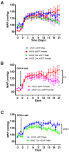
No sex differences in the severity of hypertension. Renin-a–floxed mice were injected into the SFO with either AAV2-eGFP or AAV2-Cre-eGFP. BP was monitored by telemetry for 3 days before and 21 days after DOCA-salt treatment. Continuous mean arterial pressure (MAP) recording before and during 21 days of DOCA-salt treatment, separated into (A) all males and females (n = 15–16 mice/group), (B) females only (n = 5 mice/group), and (C) males only (n = 10–11 mice/group). ** p < 0.01, **** p < 0.0001 versus AAV2-eGFP DOCA-salt (two-way ANOVA with Fisher’s LSD test).
Figure 8
Figure 8
Renin-a deletion in the SFO improves autonomic function in DOCA-salt hypertensive mice. Autonomic function was assessed by intraperitoneal injection of the ganglionic blocker chlorisondamine (6 mg/kg), muscarinic receptor blocker methylatropine (1 mg/kg), or β-adrenergic receptor blocker propranolol (5 mg/kg). (A) Reduction in BP response to chlorisondamine, indicative of the neurogenic contribution to BP (n = 9–13 mice/group). (B) Increase in HR response to methylatropine, indicative of cardiac parasympathetic tone (n = 4–6 mice/group). (C) Reduction in HR response to propranolol, indicative of cardiac sympathetic tone (n = 5–9 mice/group). ** p < 0.01, *** p < 0.001, **** p < 0.0001 versus baseline; # p < 0.05, ## p < 0.01 versus AAV2-eGFP DOCA-salt (one-way ANOVA with Fisher’s LSD test).
Figure 9
Figure 9
Renin-a deletion in the SFO prevents upregulation of NOX2 during DOCA-hypertension. mRNA expression of target genes measured in SFO samples from DOCA-Salt or Sham mice on days 7 and 14 of treatment. (A) Quantitative PCR analysis of mRNA expression of Ang II receptors (AT1aR and AT2R) and (B) NAD(P)H oxidases (NOX2 and NOX4). (C) Mechanistic hypothesis for the effect of SFO ablation of renin-a on salt-sensitive hypertension (illustration created using Biorender); pathways in dotted gray square were not directly tested in this study. * p < 0.05, ** p < 0.01 versus baseline AAV2-eGFP Sham; ## p < 0.01 versus AAV2-eGFP DOCA-salt (one-way ANOVA with Fisher’s LSD test). Abbreviations: AT1aR, angiotensin II type 1a receptor; AT2R, angiotensin II type 2 receptor; NOX2, NADPH oxidase isoform 2; NOX4, NADPH oxidase isoform 4; SFO, subfornical organ.

References

    1. Worker C.J., Li W., Feng C.Y., Souza L.A.C., Gayban A.J.B., Cooper S.G., Afrin S., Romanick S., Ferguson B.S., Feng Earley Y. The Neuronal (Pro)renin Receptor and Astrocyte Inflammation in the Central Regulation of Blood Pressure and Blood Glucose in Mice Fed a High-Fat Diet. Am. J. Physiol. Endocrinol. Metab. 2020;318:E765–E778. doi: 10.1152/ajpendo.00406.2019. - DOI - PMC - PubMed
    1. Almeida L.F., Tofteng S.S., Madsen K., Jensen B.L. Role of the renin-angiotensin system in kidney development and programming of adult blood pressure. Clin. Sci. 2020;134:641–656. doi: 10.1042/CS20190765. - DOI - PubMed
    1. Nakagawa P., Gomez J., Grobe J.L., Sigmund C.D. The Renin-Angiotensin System in the Central Nervous System and Its Role in Blood Pressure Regulation. Curr. Hypertens. Rep. 2020;22:7. doi: 10.1007/s11906-019-1011-2. - DOI - PMC - PubMed
    1. Dzau V.J. Circulating versus local renin-angiotensin system in cardiovascular homeostasis. Circulation. 1988;77:I4–I13. - PubMed
    1. Paul M., Poyan Mehr A., Kreutz R. Physiology of local renin-angiotensin systems. Physiol. Rev. 2006;86:747–803. doi: 10.1152/physrev.00036.2005. - DOI - PubMed

Publication types