. 2022 Aug 31;12(9):1213.
doi: 10.3390/biom12091213.

Role of Arginase-II in Podocyte Injury under Hypoxic Conditions

Affiliations

Role of Arginase-II in Podocyte Injury under Hypoxic Conditions

Zhilong Ren et al. Biomolecules. .

Abstract

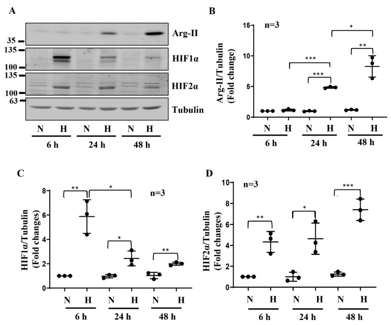
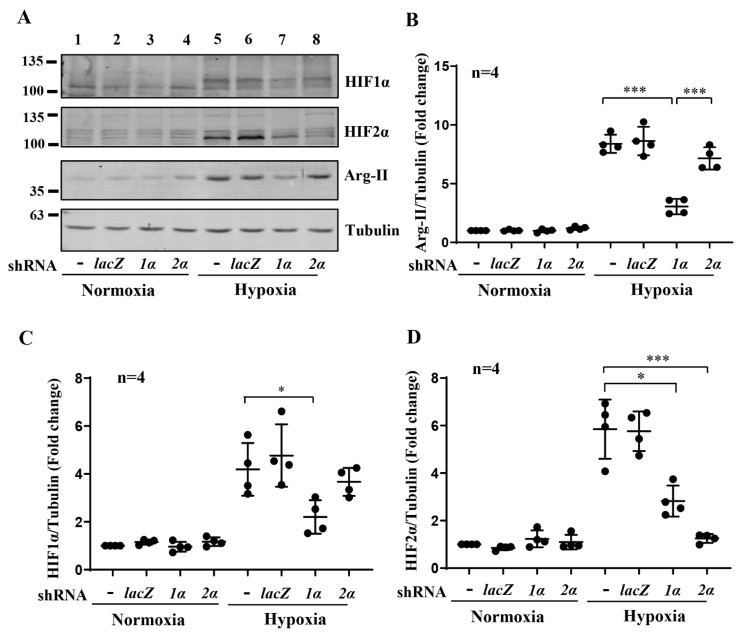
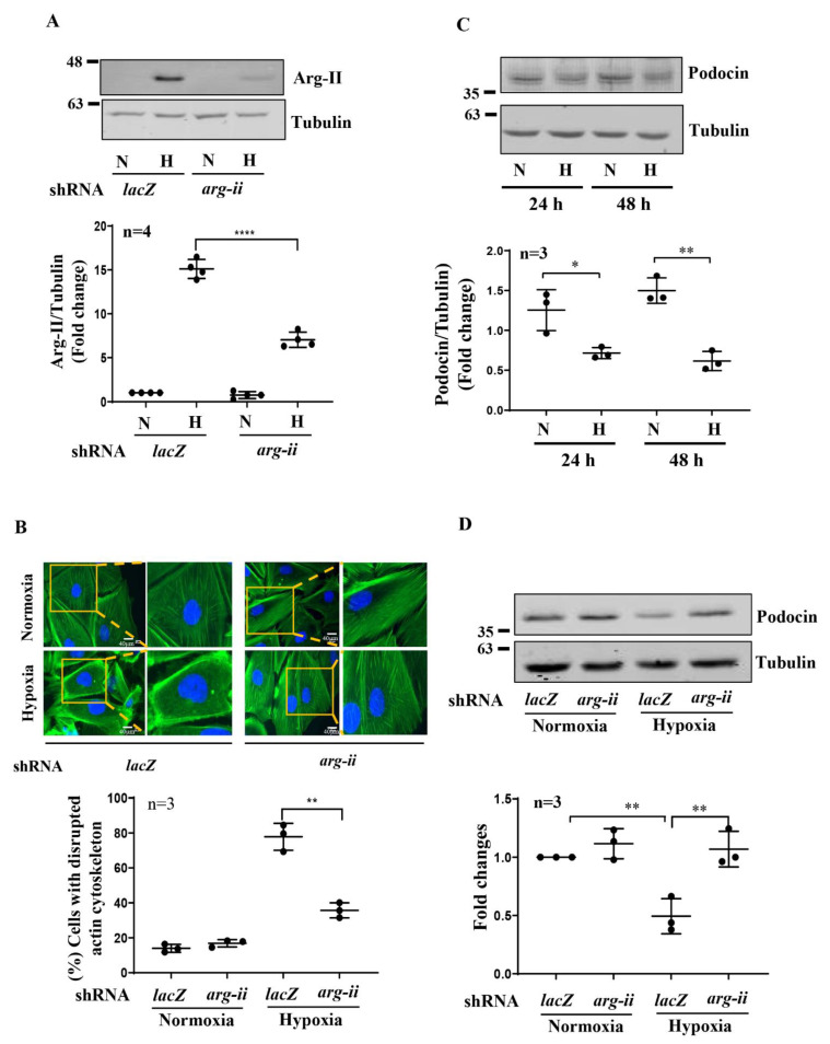
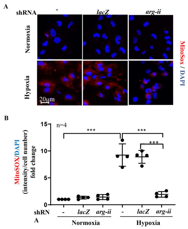
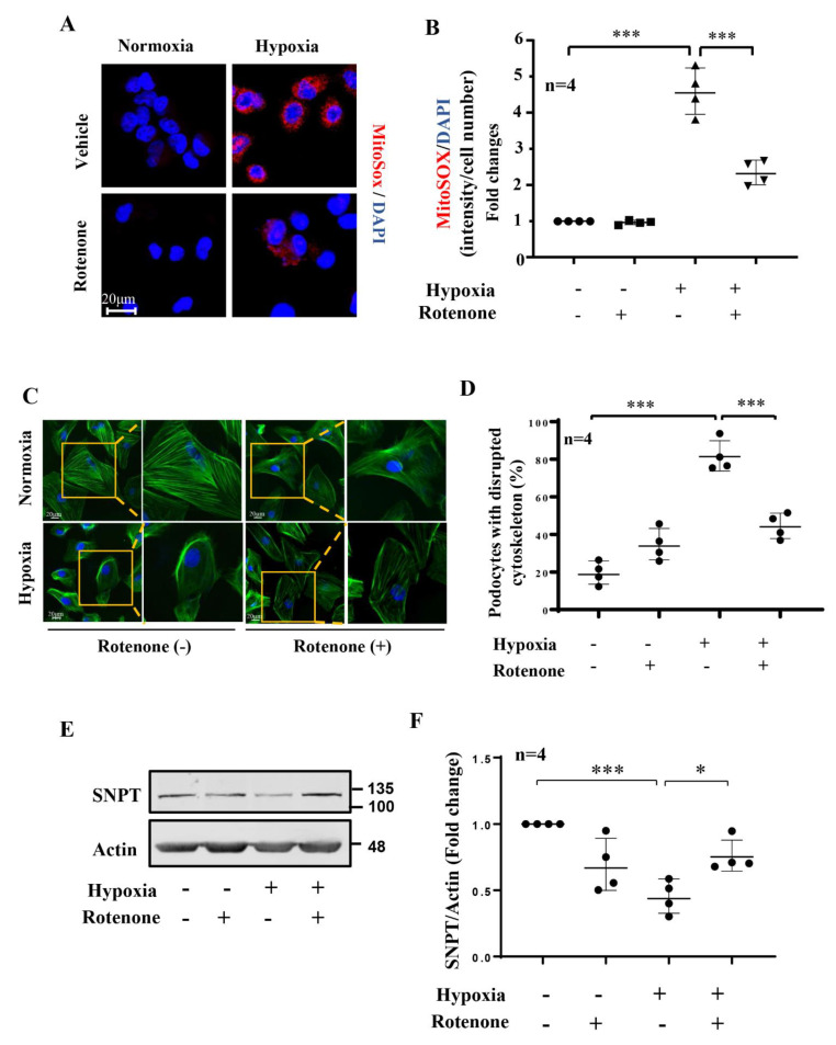
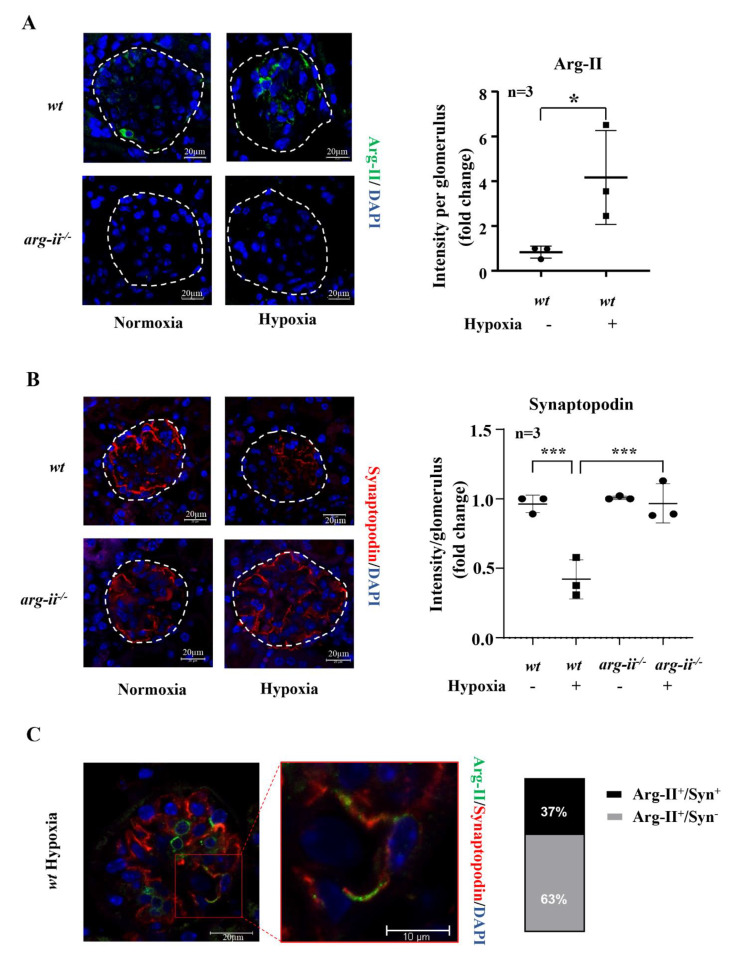
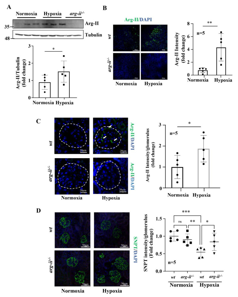
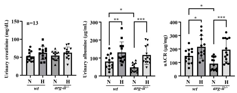
Hypoxia plays a crucial role in acute and chronic renal injury, which is attributable to renal tubular and glomerular cell damage. Some studies provide evidence that hypoxia-dependent upregulation of the mitochondrial enzyme arginase type-II (Arg-II) in tubular cells promotes renal tubular injury. It is, however, not known whether Arg-II is also expressed in glomerular cells, particularly podocytes under hypoxic conditions, contributing to hypoxia-induced podocyte injury. The effects of hypoxia on human podocyte cells (AB8/13) in cultures and on isolated kidneys from wild-type (wt) and arg-ii gene-deficient (arg-ii-/-) mice ex vivo, as well as on mice of the two genotypes in vivo, were investigated, respectively. We found that the Arg-II levels were enhanced in cultured podocytes in a time-dependent manner over 48 h, which was dependent on the stabilization of hypoxia-inducible factor 1α (HIF1α). Moreover, a hypoxia-induced derangement of cellular actin cytoskeletal fibers, a decrease in podocin, and an increase in mitochondrial ROS (mtROS) generation-as measured by MitoSOX-were inhibited by adenoviral-mediated arg-ii gene silencing. These effects of hypoxia on podocyte injury were mimicked by the HIFα stabilizing drug DMOG, which inhibits prolyl hydroxylases (PHD), the enzymes involved in HIFα degradation. The silencing of arg-ii prevented the detrimental effects of DMOG on podocytes. Furthermore, the inhibition of mtROS generation by rotenone-the inhibitor of respiration chain complex-I-recapitulated the protective effects of arg-ii silencing on podocytes under hypoxic conditions. Moreover, the ex vivo experiments with isolated kidney tissues and the in vivo experiments with mice exposed to hypoxic conditions showed increased Arg-II levels in podocytes and decreased podocyte markers regarding synaptopodin in wt mice but not in arg-ii-/- mice. While age-associated albuminuria was reduced in the arg-ii-/- mice, the hypoxia-induced increase in albuminuria was, however, not significantly affected in the arg-ii-/-. Our study demonstrates that Arg-II in podocytes promotes cell injury. Arg-ii ablation seems insufficient to protect mice in vivo against a hypoxia-induced increase in albuminuria, but it does reduce albuminuria in aging.

Keywords: HIF; ROS; arginase; hypoxia; podocytes.


Conflict of interest statement

The authors declare no competing interests.

Figures

Figure 1
Figure 1
Hypoxia enhances Arg-II levels and HIFs in human podocytes. (A) Representative immunoblotting results of the protein levels of Arg-II, HIF1α, and HIF2α in different groups of podocytes. Tubulin serves as the loading control. (BD) Quantification of the signals of Arg-II, HIF1α, and HIF2α, respectively. * p < 0.05, ** p < 0.01, *** p < 0.001 between the indicated groups. n = 3; N: normoxia; H: hypoxia, h = hours.
Figure 2
Figure 2
Role of HIFs in Arg-II upregulation by hypoxia in human podocytes. (A) Representative immunoblotting results of the protein levels of Arg-II, HIF1α, and HIF2α in different groups of podocytes. Tubulin serves as the loading control. (B–D) Quantification of the signals of Arg-II, HIF1α, and HIF2α, respectively. Lanes 1 and 5: wild-type podocytes AB8/13; Lanes 2 and 6: podocytes transduced with control rAd/U6-lacZshRNA; Lanes 3 and 7: podocytes transduced with rAd/U6-hif1αshRNA (1α); Lanes 4 and 8: podocytes transduced with rAd/U6-hif2αshRNA (2α). * p < 0.05, *** p < 0.001 between the indicated groups. n = 4.
Figure 3
Figure 3
Silencing arg-ii in human podocytes attenuates hypoxia-induced cytoskeleton actin fiber derangement. (A) Representative immunoblotting results of Arg-II protein levels in different groups of podocytes. Tubulin serves as the loading control; n = 4. (B) Representative images showing phalloidin staining of cytoskeleton actin fibers in different groups of podocytes. Nucleoli were stained with DAPI (blue). Inserts illustrate a cell that represents the typical changes in cytoskeletal fibers under the indicated conditions. The graphics below show the quantification of podocytes with a disrupted cytoskeleton. n = 3. (C) Representative immunoblotting results show decreased podocin protein levels under hypoxic conditions; Tubulin serves as the loading control. The graphics below show the quantification of podocin signals. (D) Representative immunoblotting results show protective effects of arg-ii silencing on a hypoxia-induced decrease in podocin protein levels in podocytes. Tubulin serves as the loading control. The graphics below show the quantification of podocin signals. N: normoxia; H: hypoxia. * p < 0.05, ** p < 0.01, **** p < 0.001 between the indicated groups. n = 3.
Figure 4
Figure 4
Silencing arg-ii reduces hypoxia-enhanced mitochondrial ROS production in podocytes. MitoSOX Red reagent staining was used to analyze mitochondrial ROS production. -: wild-type podocytes; shRNA-lacZ: podocytes transduced with rAd/U6-lacZshRNA as controls; shRNA-arg-ii: podocytes transduced with rAd/U6-arg-iishRNA. (A). Representative images of MitoSOX staining; (B). Quantification of relative fluorescence fold-change in different groups of podocytes. *** p < 0.001 between the indicated groups. n = 4.
Figure 5
Figure 5
Role of mitochondrial ROS on hypoxia-induced podocyte injury. The cells were pretreated with or without rotenone (2 μmol/L) for 1 h and then incubated under normoxia or hypoxia (2% O2) conditions for 24 h. (A). Representative images show MitoSOX staining in human podocytes under different conditions. Nucleoli were stained with DAPI (blue). (B). Quantification of relative fluorescence fold-change in different groups of podocytes. (C). Representative images show phalloidin staining of cytoskeletal actin fibers in human podocytes under different conditions. Nucleoli were stained with DAPI (blue). Inserts illustrate a cell that represents the typical changes in cytoskeletal fibers under the indicated conditions. (D). Quantification of the podocytes with the disrupted actin cytoskeleton. (E). Representative immunoblotting results of the protein levels of synaptopodin (SNPT) in different groups of podocytes; β-actin serves as the loading control. (F). Quantification of synaptopodin signals. * p < 0.05, *** p < 0.001 between the indicated groups. n = 4.
Figure 6
Figure 6
Arg-ii ablation prevents hypoxia-induced synaptopodin reduction in glomeruli ex vivo. Confocal immunofluorescence staining of (A) Arg-II, (B) synaptopodin and (C) co-localization of Arg-II (green) and synaptopodin (red) with quantification of Arg-II+/synaptopodin+ and Arg-II+/synaptopodin signals in isolated kidneys from wt and arg-ii−/− mice that were exposed to normoxia or hypoxia conditions (1% O2, 24 h) ex vivo. Nucleoli were stained with DAPI (blue). n = 3, * p < 0.05, *** p < 0.001 between the indicated groups.
Figure 7
Figure 7
Arg-ii ablation prevents hypoxia-induced reduction in synaptopodin in vivo. (A). Representative immunoblotting shows Arg-II levels and relative quantification of Arg-II levels in wt under normoxic and hypoxic conditions. Tubulin serves as the loading control. (B) Representative images show Arg-II protein expression and quantification in proximal tubular cells (PTCs). (C) Confocal immunofluorescence staining shows Arg-II protein expression and quantification in glomeruli; (D). Confocal immunofluorescence staining of synaptopodin (SNPT) and quantification in wt and arg-ii−/− mouse kidneys (n = 5 for each group). Nucleoli were stained with DAPI (blue). * p < 0.05, ** p < 0.01, *** p < 0.001 between the indicated groups.
Figure 8
Figure 8
Effect of arg-ii ablation on albuminuria. Urinary creatinine and albuminuria were assessed in 20 to 24 months old mice exposed to normoxia controls or hypoxia (8% O2 for 24 h). The urinary albumin/creatinine ratio (uACR) was evaluated in wt and arg-ii−/− animals. n = 13 in each group. * p < 0.05, ** p < 0.01, *** p < 0.005 between the indicated groups.

References

    1. Pavenstadt H., Kriz W., Kretzler M. Cell biology of the glomerular podocyte. Physiol. Rev. 2003;83:253–307. doi: 10.1152/physrev.00020.2002. - DOI - PubMed
    1. Nagata M. Podocyte injury and its consequences. Kidney Int. 2016;89:1221–1230. doi: 10.1016/j.kint.2016.01.012. - DOI - PubMed
    1. Shankland S.J., Wang Y., Shaw A.S., Vaughan J.C., Pippin J.W., Wessely O. Podocyte Aging: Why and How Getting Old Matters. J. Am. Soc. Nephrol. 2021;32:2697–2713. doi: 10.1681/ASN.2021050614. - DOI - PMC - PubMed
    1. O’Connor P.M. Renal oxygen delivery: Matching delivery to metabolic demand. Clin. Exp. Pharmacol. Physiol. 2006;33:961–967. doi: 10.1111/j.1440-1681.2006.04475.x. - DOI - PubMed
    1. Faivre A., Scholz C.C., de Seigneux S. Hypoxia in chronic kidney disease: Towards a paradigm shift? Nephrol. Dial. Transplant. 2020;36:1782–1790. doi: 10.1093/ndt/gfaa091. - DOI - PubMed

Publication types