. 2022 Sep 7;11(18):2797.
doi: 10.3390/cells11182797.

MicroRNA-4516 in Urinary Exosomes as a Biomarker of Premature Ovarian Insufficiency

Affiliations

MicroRNA-4516 in Urinary Exosomes as a Biomarker of Premature Ovarian Insufficiency

Zobia Umair et al. Cells. .

Abstract

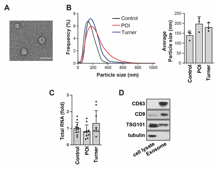
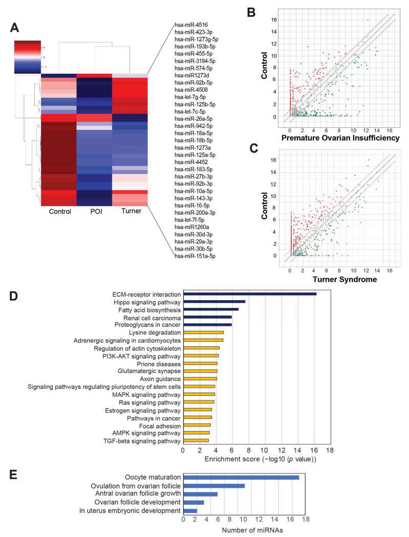
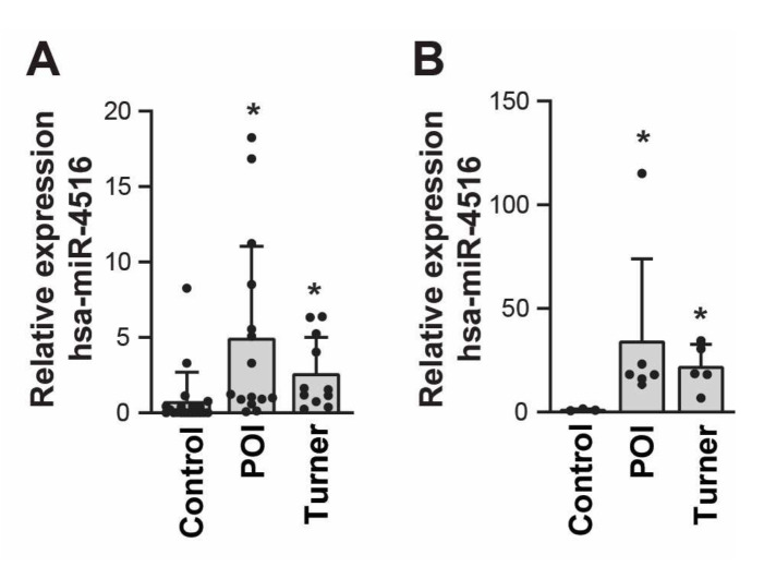
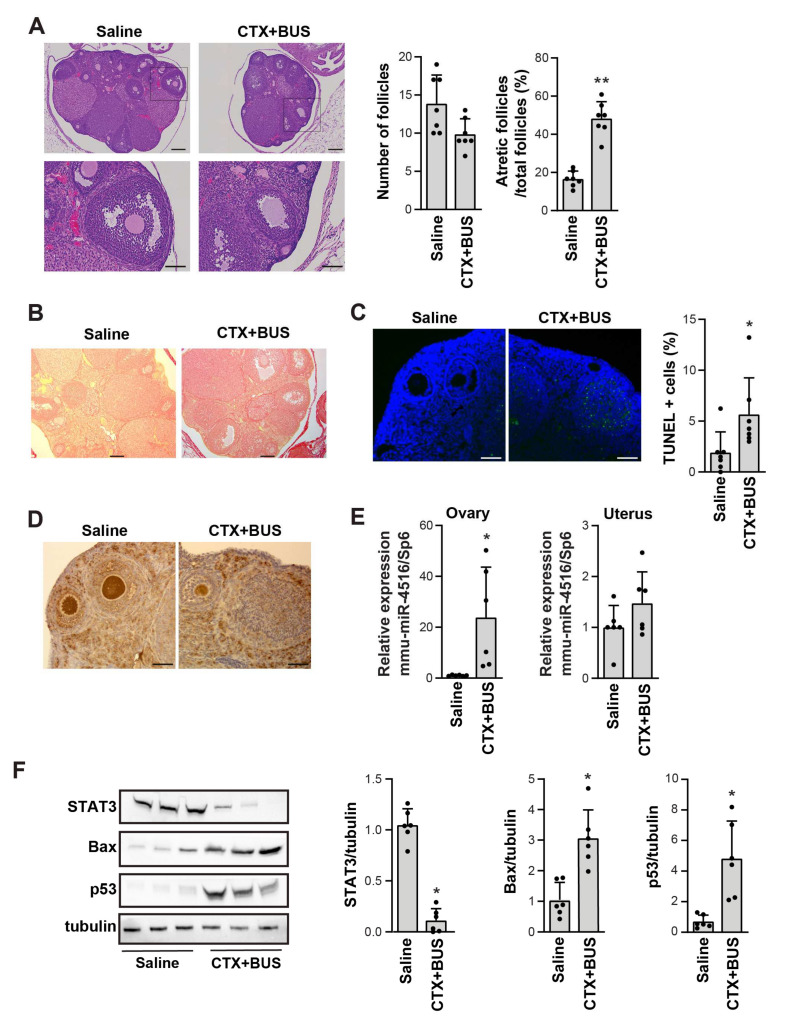
Premature ovarian insufficiency (POI) is a typical disorder of amenorrhea that lasts for a minimum of four months in women < 40 years old and is typically characterized by reduced estrogen levels and elevated serum concentrations of follicle-stimulating hormone. We collected urine samples from two participant cohorts from Gil Hospital of Gachon University (Incheon, Korea): a sequencing cohort of 19 participants (seven patients with POI (POI patients without Turner syndrome), seven patients with Turner syndrome (POI patients with Turner syndrome), and five control individuals (age-matched controls with confirmed ovarian sufficiency)) and a validation cohort of 46 participants (15 patients with POI, 11 patients with Turner syndrome, and 20 control individuals). Among differentially expressed miRNAs, hsa-miR-4516 was significantly upregulated in patients with POI in both cohorts, independent of the presence of Turner syndrome. Moreover, the upregulation of miR-4516 was confirmed in the ovary-but not in the uterus-of a cyclophosphamide and busulfan-induced POI mouse model. This was accompanied by a decrease in STAT3 protein level, a predicted target of miR-4516, via miRTarBase2020. Our study provides compelling evidence that miR-4516 is highly expressed in patients with POI and POI mouse models, suggesting that miR-4516 is a diagnostic marker of POI.

Keywords: hsa-miR-4516; micro-RNA; premature ovarian insufficiency; turner syndrome; urinary exosome.


Conflict of interest statement

The authors declare no conflict of interest.

Figures

Figure 1
Figure 1
Characterization of exosomes isolated from the urine. (A) Phenotype of urinary exosomes detected via transmission electron microscopy. Scale bar: 100 nm. (B) Frequency of isolated urinary exosomes revealed via nanoparticle tracking analysis. All average particle sizes were calculated by using three independent samples. (C) The amount of total RNA from each sample was measured by using Nanodrop 2000 (ThermoFisher Scientific); patients with premature ovarian insufficiency (POI) = 11, patients with Turner syndrome = 9, control individuals = 12. (D) Expression levels of CD63, CD9, and TSG101 in urinary exosomes were detected via Western blot analysis. Tubulin was used as a negative control.
Figure 2
Figure 2
MicroRNA (miRNA) sequencing profile of urinary exosomes from patients with premature ovarian insufficiency (POI), Turner syndrome, and control individuals. (A) Heatmap with hierarchical clustering of 33 differentially expressed miRNAs (p < 0.05). Red and green colors represent increased or reduced expressions across samples, respectively. (B,C) Scatterplots of miRNA profiles among patients with POI versus the control (B) and patients with Turner syndrome versus the control (C). (D) Top 20 statistically significant enriched pathways in the Kyoto Encyclopedia of Genes and Genomes (KEGG) analyses of the genes targeted by the 33 differentially expressed miRNAs. (E) Gene category analysis for the POI-related target genes of the 33 differentially expressed miRNAs.
Figure 3
Figure 3
Relative expression of hsa-miR-4516 in the validation cohorts. (A) Relative expression of hsa-miR-4516 in the validation cohort, using qRT-PCR (patients with premature ovarian insufficiency (POI) = 15, patients with Turner syndrome = 11, and control individuals = 20). Relative miRNA expression was normalized by using UniSp6 as an internal control. (B) Relative expression of hsa-miR-4516, using real-time Taqman analysis, was normalized by ath-miR-159a as an endogenous control (patients with premature ovarian insufficiency (POI) = 6, patients with Turner syndrome = 5, and control individuals = 3). The p-value was evaluated by comparison to the control group. Asterisks (*) indicate significant differences (p < 0.05) when compared to the control group.
Figure 4
Figure 4
Increase in miR-4516 expression in a chemotherapy-induced premature ovarian insufficiency (POI) mouse model. (A) Representative hematoxylin-and-eosin-stained images of the ovaries from saline- and CTX+BUS-treated mice (n = 7). Scale bar = 200 μm in the upper panel and 80 μm in the lower panel. (B) Representative images of Sirius red staining in the ovaries from saline- and CTX+BUS-treated mice. Scale bar = 200 μm. (C) Representative images of the TUNEL assay in the ovaries from saline- and CTX+BUS-treated mice. Scale bar = 80 μm. TUNEL-positive cells were counted by using ImageJ software (n = 7). (D) Representative immunohistochemistry images of Ki67 expression in the ovaries from saline- and CTX+BUS-treated mice. Scale bar = 80 μm. (E) Relative expression of mmu-miR-4516 in the ovaries and uteri from the saline- and CTX+BUS-treated mice were analyzed by using qRT-PCR. Relative miRNA expression was normalized by using UniSp6 as an internal control (n = 6). (F) Western blot analysis of STAT3, Bax, and p53 expression in the ovaries of saline- and CTX+BUS-treated mice. The relative protein level in (F) was normalized to tubulin levels (mean ± SD; n = 6). Asterisks (*, **) indicate significant differences (p < 0.05, p < 0.01, respectively) when compared to saline-injected mice.

References

    1. Rudnicka E., Kruszewska J., Klicka K., Kowalczyk J., Grymowicz M., Skorska J., Pieta W., Smolarczyk R. Premature ovarian insufficiency-aetiopathology, epidemiology, and diagnostic evaluation. Prz. Menopauzalny. 2018;17:105–108. doi: 10.5114/pm.2018.78550. - DOI - PMC - PubMed
    1. Haller-Kikkatalo K., Uibo R., Kurg A., Salumets A. The prevalence and phenotypic characteristics of spontaneous premature ovarian failure: A general population registry-based study. Hum. Reprod. 2015;30:1229–1238. doi: 10.1093/humrep/dev021. - DOI - PubMed
    1. Roeters van Lennep J.E., Heida K.Y., Bots M.L., Hoek A., collaborators of the Dutch Multidisciplinary Guideline Development Group on Cardiovascular Risk Management after Reproductive, D Cardiovascular disease risk in women with premature ovarian insufficiency: A systematic review and meta-analysis. Eur. J. Prev. Cardiol. 2016;23:178–186. doi: 10.1177/2047487314556004. - DOI - PubMed
    1. Asli I.N., Fallahian M., Seddigh H.R., Javadi H., Baharfar N., Assadi M. Evaluation of bone mineral density in premature ovarian failure. Hell. J. Nucl. Med. 2010;13:261–263. - PubMed
    1. Bove R., Secor E., Chibnik L.B., Barnes L.L., Schneider J.A., Bennett D.A., De Jager P.L. Age at surgical menopause influences cognitive decline and Alzheimer pathology in older women. Neurology. 2014;82:222–229. doi: 10.1212/WNL.0000000000000033. - DOI - PMC - PubMed

Publication types