. 2022 Sep 9;11(18):2819.
doi: 10.3390/cells11182819.

Proteomic Analysis of Retinal Mitochondria-Associated ER Membranes Identified Novel Proteins of Retinal Degeneration in Long-Term Diabetes

Affiliations

Proteomic Analysis of Retinal Mitochondria-Associated ER Membranes Identified Novel Proteins of Retinal Degeneration in Long-Term Diabetes

Joshua J Wang et al. Cells. .

Abstract

The mitochondria-associated endoplasmic reticulum (ER) membrane (MAM) is the physical contact site between the ER and the mitochondria and plays a vital role in the regulation of calcium signaling, bioenergetics, and inflammation. Disturbances in these processes and dysregulation of the ER and mitochondrial homeostasis contribute to the pathogenesis of diabetic retinopathy (DR). However, few studies have examined the impact of diabetes on the retinal MAM and its implication in DR pathogenesis. In the present study, we investigated the proteomic changes in retinal MAM from Long Evans rats with streptozotocin-induced long-term Type 1 diabetes. Furthermore, we performed in-depth bioinformatic analysis to identify key MAM proteins and pathways that are potentially implicated in retinal inflammation, angiogenesis, and neurodegeneration. A total of 2664 unique proteins were quantified using IonStar proteomics-pipeline in rat retinal MAM, among which 179 proteins showed significant changes in diabetes. Functional annotation revealed that the 179 proteins are involved in important biological processes such as cell survival, inflammatory response, and cellular maintenance, as well as multiple disease-relevant signaling pathways, e.g., integrin signaling, leukocyte extravasation, PPAR, PTEN, and RhoGDI signaling. Our study provides comprehensive information on MAM protein changes in diabetic retinas, which is helpful for understanding the mechanisms of metabolic dysfunction and retinal cell injury in DR.

Keywords: Type 1 diabetes; diabetic retinopathy; mitochondria-associated ER membrane; proteomic; retinal degeneration.


Conflict of interest statement

The authors declare no conflict of interest.

Figures
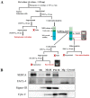
Figure 1
Figure 1
Schematic flowchart of the MAM isolation protocol and Western blot validation of MAM purity. (A) MAM was isolated from rat retina via homogenization, differential centrifugations, and a self-forming Percoll gradient centrifugation. ER, crude mitochondria, and pure mitochondria were also isolated following the multiple centrifugation steps. (B) Western blot analysis of MAM, ER and/or mitochondrial markers from retinal MAM fractions, ER, cytosol, and mitochondrial isolates. ER: endoplasmic reticulum, Mc: crude mitochondria, MAM: ER mitochondria-associated membrane, Pel-M: pellet after centrifugation of crude MAM, Mp: pure mitochondria, SERCA: Sarco/endoplasmic reticulum Ca2+-ATPase, FACL-4: Acyl-CoA synthetase 4, Sigma-1R: Sigma-1 receptor, Cyto C: cytochrome C.
Figure 2
Figure 2
Subcellular localization and biological relevance of significantly altered MAM proteins in Type 1 diabetic rat retinas identified by proteomics. (A) Organelle association of the 179 altered MAM proteins based on cross-referenced annotation. (B) Percentages of molecule types, according to GO analysis from available database information. MAM: Mitochondria-associated ER membrane, GO: Gene ontology.
Figure 3
Figure 3
Hierarchical bar graph of key biological processes pertaining to significantly altered MAM proteins in Type 1 diabetic rat retinas. Biological processes of the 179 altered MAM proteins are classified as one of four categories of cellular activities or “other” according to GO analysis. MAM: Mitochondria-associated ER membrane, GO: Gene ontology.
Figure 4
Figure 4
Bar graph of affected pathophysiologic processes that contribute to DR in the MAM of Type 1 diabetic rat retinas. MAM proteins are classified as up- or down-regulated as well as categorized into 20 biological processes that are labeled as functionally increased, decreased, or generally affected. DR: diabetic retinopathy, MAM: Mitochondria-associated ER membrane.
Figure 5
Figure 5
General and pathophysiology-specific heat maps of significantly altered MAM proteins in Type 1 diabetic rat retinas. MAM proteins that were most affected by fold-change ratio were selected among the 179 altered MAM proteins in the Type 1 diabetic condition. Proteins with a p-value of less than 0.05 were considered significant. p-values for each altered protein are shown on the right of each map. NDM: non-diabetes mellitus, DM: diabetes mellitus, MAM: Mitochondria-associated ER membrane.
Figure 6
Figure 6
Interaction map depicting MAM proteins and their involvement with major inflammatory mediators. MAM proteins identified to have interactions with inflammatory mediators were selected from the 179 altered MAM proteins in the Type 1 diabetic condition. Numerous interactions were specifically identified with Interleukin-1β (IL-1β), NF-kappa-B inhibitor alpha (NF-κBIA), Forkhead box protein O1 (FOXO1), endothelin-1 (EDN1), and c-Jun (JUN). The functional networks were generated through IPA. Legend in the box displays molecules and function symbol, types, and colors. Lighter shades of red, green, and blue represent decreased intensities of up-regulation, down-regulation, and inhibition, respectively. MAM: Mitochondria-associated ER membrane, IPA: Ingenuity pathway analysis.
Figure 7
Figure 7
Interaction maps of MAM proteins and their multiple protein-protein associations with pathways of retinal degeneration. MAM proteins commonly identified to have protein-protein interactions with retinal degeneration processes were selected from the 179 altered MAM proteins in the Type 1 diabetic condition. (A) Eleven MAM proteins were found to have strong associations with SYVN1 as well as ties to glucose metabolism disorder and processes of molecular transport and endocytosis. (B) Five MAM proteins were found to have strong associations with STAT4 and multiple key intracellular processes. (C) Seventeen MAM proteins have common connections with processes of photoreceptor and retinal degeneration. (D) Ten MAM proteins each have associations with FOXO1 as well as intricate associations with glucose synthesis and metabolism, hyperlipidemia, and retinal neovascularization. The functional networks were generated through IPA. Legend displays molecules and function symbol types and colors. Lighter shades of red, green, and blue represent decreased intensities of up-regulation, down-regulation, and inhibition, respectively.
Figure 8
Figure 8
Interaction maps of MAM proteins and associated eye pathologies and canonical DR-related signaling pathways. MAM proteins with notable associations with specific signaling pathways and eye pathology underlying DR were selected from the 179 altered MAM proteins in the Type 1 diabetic condition. (A) Eight crystallin proteins and VCAM1 were notably identified as having associations with MAF. VCAM1 and several specific crystallin proteins (CRYAB, CRYAA/CRYAA2) were found to have numerous ties to the onset of various eye pathologies. (B) LGALS3 is a key MAM protein that is associated with processes of retinal degeneration via multiple specific protein-protein interactions. The functional networks were generated through IPA. Figure legend displays molecules and function symbol types and colors. Lighter shades of red, green, and blue represent decreased intensities of up-regulation, down-regulation, and inhibition, respectively. MAM: Mitochondria-associated ER membrane, IPA: Ingenuity pathway analysis, VCAM-1: Vascular cell adhesion molecule 1, MAF: MAF bZIP transcription factor, CRYAA: Crystallin alpha A, CRYAA: Crystallin alpha B, CRYAA2: Crystallin alpha A2, LGAL3: Galectin 3.

References

    1. Fong D.S., Aiello L., Gardner T.W., King G.L., Blankenship G., Cavallerano J.D., Ferris F.L., 3rd, Klein R. American Diabetes A: Retinopathy in diabetes. Diabetes Care. 2004;27((Suppl. 1)):S84–S87. doi: 10.2337/diacare.27.2007.S84. - DOI - PubMed
    1. Reis A., Mateus C., Melo P., Figueira J., Cunha-Vaz J., Castelo-Branco M. Pure neuroretinal dysfunction in diabetic retinopathy occurring prior to endothelial and vascular damage. Acta Ophthalmol. 2013;91:S252. doi: 10.1111/j.1755-3768.2013.1662.x. - DOI
    1. Solomon S.D., Chew E., Duh E.J., Sobrin L., Sun J.K., VanderBeek B.L., Wykoff C.C., Gardner T.W. Diabetic Retinopathy: A Position Statement by the American Diabetes Association. Diabetes Care. 2017;40:412–418. doi: 10.2337/dc16-2641. - DOI - PMC - PubMed
    1. Simo R., Stitt A.W., Gardner T.W. Neurodegeneration in diabetic retinopathy: Does it really matter? Diabetologia. 2018;61:1902–1912. doi: 10.1007/s00125-018-4692-1. - DOI - PMC - PubMed
    1. Lee V.K., Hosking B.M., Holeniewska J., Kubala E.C., Lundh von Leithner P., Gardner P.J., Foxton R.H., Shima D.T. BTBR ob/ob mouse model of type 2 diabetes exhibits early loss of retinal function and retinal inflammation followed by late vascular changes. Diabetologia. 2018;61:2422–2432. doi: 10.1007/s00125-018-4696-x. - DOI - PMC - PubMed

Publication types

Substances