. 2022 Sep 15;11(18):2882.
doi: 10.3390/cells11182882.

7-Ketocholesterol Induces Oxiapoptophagy and Inhibits Osteogenic Differentiation in MC3T3-E1 Cells

Affiliations

7-Ketocholesterol Induces Oxiapoptophagy and Inhibits Osteogenic Differentiation in MC3T3-E1 Cells

Jing Ouyang et al. Cells. .

Abstract

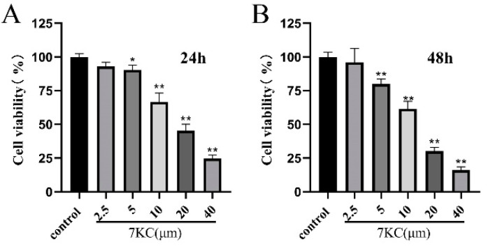
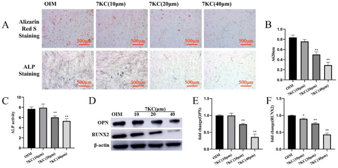
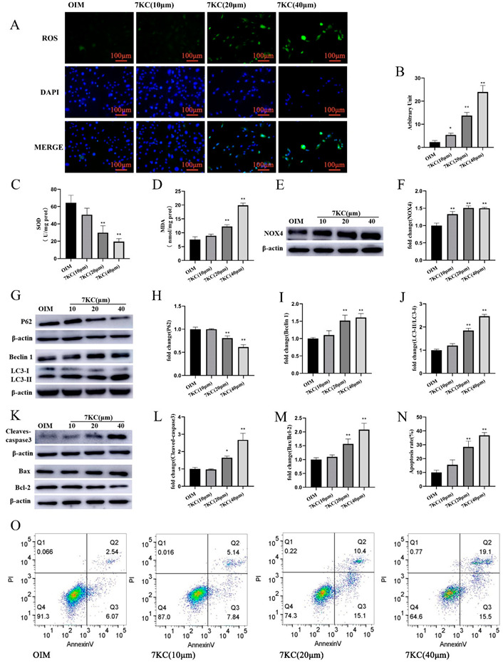
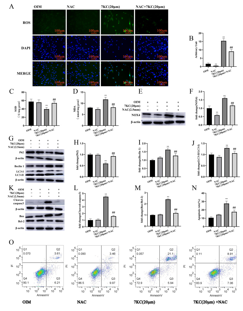
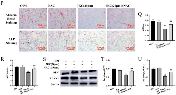
7-Ketocholesterol (7KC) is one of the oxysterols produced by the auto-oxidation of cholesterol during the dysregulation of cholesterol metabolism which has been implicated in the pathological development of osteoporosis (OP). Oxiapoptophagy involving oxidative stress, autophagy, and apoptosis can be induced by 7KC. However, whether 7KC produces negative effects on MC3T3-E1 cells by stimulating oxiapoptophagy is still unclear. In the current study, 7KC was found to significantly decrease the cell viability of MC3T3-E1 cells in a concentration-dependent manner. In addition, 7KC decreased ALP staining and mineralization and down-regulated the protein expression of OPN and RUNX2, inhibiting osteogenic differentiation. 7KC significantly stimulated oxidation and induced autophagy and apoptosis in the cultured MC3T3-E1 cells. Pretreatment with the anti-oxidant acetylcysteine (NAC) could effectively decrease NOX4 and MDA production, enhance SOD activity, ameliorate the expression of autophagy-related factors, decrease apoptotic protein expression, and increase ALP, OPN, and RUNX2 expression, compromising 7KC-induced oxiapoptophagy and osteogenic differentiation inhibition in MC3T3-E1 cells. In summary, 7KC may induce oxiapoptophagy and inhibit osteogenic differentiation in the pathological development of OP.

Keywords: 7-Ketocholesterol; osteogenic differentiation; osteoporosis; oxiapoptophagy; oxidative stress.


Conflict of interest statement

The authors declare no conflict of interest.

Figures

Figure 1
Figure 1
7KC inhibited MC3T3-E1 cell viability. (A,B) MC3T3-E1 cells were treated with 7KC (0, 2.5, 5, 10, 20, or 40 μM) for 24 h and 48 h. * p < 0.05; ** p < 0.01.
Figure 2
Figure 2
7KC inhibited the osteogenic differentiation of MC3T3-E1 cells. MC3T3-E1 cells were treated with 7KC (0, 10, 20, or 40 μM). (AC) ALP activity detection and the mineral assays were performed (×50 magnification); ALP activity detection and quantitative analysis of the mineralized area. The protein expressions of OPN (D,E) and RUNX2 (D,F) were analyzed by Western blot. Data were analyzed and compared with the OIM group. * p < 0.05; ** p < 0.01. OIM, osteogenic induction medium.
Figure 3
Figure 3
7KC regulated oxiapoptophagy in MC3T3-E1 cells. (A) Cells were prepared for staining with DCFH-DA and DAPI, respectively, and analyzed by a fluorescence microscope (×200 magnification). (B) Quantitative analysis of ROS production. (C,D) SOD activity detection, and MDA level measurement were conducted using the kits. (E,F) The protein expression of NOX4 was analyzed by Western blot. (GJ) The protein expression of LC3I/II, Beclin1, and P62 was detected. (KM) The protein expression of Bax/Bcl-2 and cleaved caspase-3 was measured. (N,O) Apoptosis analysis was conducted by flow cytometry. Data were analyzed and compared with the OIM group. * p < 0.05; ** p < 0.01. OIM, osteogenic induction medium; NC, negative control.
Figure 4
Figure 4
NAC inhibited oxiapoptophagy and differentiation in 7KC-induced MC3T3-E1 cells. (A) Cells were prepared for staining with DCFH-DA and DAPI, respectively, and analyzed by a fluorescence microscope (×200 magnification). (B) The production of ROS was analyzed by fluorescence intensity. (C,D) SOD activity detection and MDA level measurement were conducted using the kits. (E,F) The protein expression of NOX4 was analyzed by Western blot. (GJ) The protein expression of LC3I/II, Beclin1, and P62 was detected. (KM) The protein expression of Bax/Bcl-2 and cleaved caspase-3 was measured. (N,O) Apoptosis analysis was conducted by flow cytometry. (PR) ALP activity detection and the mineral assays were performed (×50 magnification) and analyzed. The protein expression of OPN (S,T) and RUNX2 (S,U) was determined by Western blotting. Data were analyzed for comparison with the OIM group. ** p < 0.01. Compared with the 7KC (20 μM) group, ## p < 0.01. OIM, osteogenic induction medium; NAC, 2.5 mM NAC.
Figure 4
Figure 4
NAC inhibited oxiapoptophagy and differentiation in 7KC-induced MC3T3-E1 cells. (A) Cells were prepared for staining with DCFH-DA and DAPI, respectively, and analyzed by a fluorescence microscope (×200 magnification). (B) The production of ROS was analyzed by fluorescence intensity. (C,D) SOD activity detection and MDA level measurement were conducted using the kits. (E,F) The protein expression of NOX4 was analyzed by Western blot. (GJ) The protein expression of LC3I/II, Beclin1, and P62 was detected. (KM) The protein expression of Bax/Bcl-2 and cleaved caspase-3 was measured. (N,O) Apoptosis analysis was conducted by flow cytometry. (PR) ALP activity detection and the mineral assays were performed (×50 magnification) and analyzed. The protein expression of OPN (S,T) and RUNX2 (S,U) was determined by Western blotting. Data were analyzed for comparison with the OIM group. ** p < 0.01. Compared with the 7KC (20 μM) group, ## p < 0.01. OIM, osteogenic induction medium; NAC, 2.5 mM NAC.

Comment in

References

    1. Eun S.Y., Cheon Y.H., Park G.D., Chung C.H., Lee C.H., Kim J.Y., Lee M.S. Anti-Osteoporosis Effects of the Eleutherococcus senticosus, Achyranthes japonica, and Atractylodes japonica Mixed Extract Fermented with Nuruk. Nutr. 2021;13:3904. doi: 10.3390/nu13113904. - DOI - PMC - PubMed
    1. Che Y., Yang J., Tang F., Wei Z., Chao Y., Li N., Li H., Wu S., Dong X. New Function of Cholesterol Oxidation Products Involved in Osteoporosis Pathogenesis. Int. J. Mol. Sci. 2022;23:2020. doi: 10.3390/ijms23042020. - DOI - PMC - PubMed
    1. Xu Z., Xie Z., Sun J., Huang S., Chen Y., Li C., Sun X., Xia B., Tian L., Guo C., et al. Gut Microbiome Reveals Specific Dysbiosis in Primary Osteoporosis. Front. Cell. Infect. Microbiol. 2020;10:160. doi: 10.3389/fcimb.2020.00160. - DOI - PMC - PubMed
    1. Yang T.L., Shen H., Liu A., Dong S.S., Zhang L., Deng F.Y., Zhao Q., Deng H.W. A road map for understanding molecular and genetic determinants of osteoporosis. Nat. Rev. Endocrinol. 2020;16:91–103. doi: 10.1038/s41574-019-0282-7. - DOI - PMC - PubMed
    1. Tian L., Yu X. Lipid metabolism disorders and bone dysfunction--interrelated and mutually regulated (review) Mol. Med. Rep. 2015;12:783–794. doi: 10.3892/mmr.2015.3472. - DOI - PMC - PubMed

Publication types

MeSH terms