. 2022 Sep 19;11(18):2926.
doi: 10.3390/cells11182926.

MARCKS as a Potential Therapeutic Target in Inflammatory Breast Cancer

Affiliations

MARCKS as a Potential Therapeutic Target in Inflammatory Breast Cancer

Maroua Manai et al. Cells. .

Abstract

Inflammatory breast cancer (IBC) is the most pro-metastatic form of breast cancer (BC). We previously demonstrated that protein overexpression of Myristoylated Alanine-Rich C Kinase Substrate (MARCKS) protein was associated with shorter survival in IBC patients. MARCKS has been associated with the PI3K/AKT pathway. MARCKS inhibitors are in development. Our objective was to investigate MARCKS, expressed preferentially in IBC that non-IBC (nIBC), as a novel potential therapeutic target for IBC. The biologic activity of MPS, a MARCKS peptide inhibitor, on cell proliferation, migration, invasion, and mammosphere formation was evaluated in IBC (SUM149 and SUM190) and nIBC (MDA-MB-231 and MCF7) cell lines, as well as its effects on protein expression in the PTEN/AKT and MAPK pathways. The prognostic relevance of MARCKS and phosphatase and tensin homolog (PTEN) protein expression as a surrogate marker of metastasis-free survival (MFS) was evaluated by immunohistochemistry (IHC) in a retrospective series of archival tumor samples derived from 180 IBC patients and 355 nIBC patients. In vitro MPS impaired cell proliferation, migration and invasion, and mammosphere formation in IBC cells. MARCKS inhibition upregulated PTEN and downregulated pAKT and pMAPK expression in IBC cells, but not in nIBC cells. By IHC, MARCKS expression and PTEN expression were negatively correlated in IBC samples and were associated with shorter MFS and longer MFS, respectively, in multivariate analysis. The combination of MARCKS-/PTEN+ protein status was associated with longer MFS in IBC patient only (p = 8.7 × 10⁻³), and mirrored the molecular profile (MARCKS-downregulated/PTEN-upregulated) of MPS-treated IBC cell lines. In conclusion, our results uncover a functional role of MARCKS implicated in IBC aggressiveness. Associated with the good-prognosis value of the MARCKS-/PTEN+ protein status that mirrors the molecular profile of MPS-treated IBC cell lines, our results suggest that MARCKS could be a potential therapeutic target in patients with MARCKS-positive IBC. Future preclinical studies using a larger panel of IBC cell lines, animal models and analysis of a larger series of clinical samples are warranted in order to validate our results.

Keywords: MARCKS; MPS treatment; PTEN; inflammatory breast cancer; mechanisms; metastasis-free survival.


Conflict of interest statement

The authors declare that they have no conflict of interest.

Figures
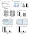
Figure 1
Figure 1
MARCKS promotes cell proliferation and motility of IBC. (A) MTT assay showed that MARCKS inhibition using 25 µM MPS peptide induced cell growth inhibition only in SUM149 compared to MDA-MB-231 cells. (B) MPS reduced colony formation of SUM149 cells, but not in MDA-MB-231: representative images (left) and box plots (right). (C) Representative images of migration in scratch/wound-healing assay to evaluate the inhibitory effect of MPS peptide on IBC vs. nIBC cell migration (left). Each confluent monolayer was wounded linearly then treated with MPS (25 µM), cell morphology and migration were observed and photographed at regular intervals, and the number of the migrated cells into the cell-free zone was calculated at 48 h compared to 0 h. (D) Similar to (C) but using the transwell migration assay. Migration was stained and measured after 24 h: representative images (top) and box plots (bottom). (E) Representative images of Matrigel invasion in chambers demonstrating the inhibitory effect of MPS in IBC cells (top) and box plots (bottom). Data were represented as mean ± SD. * for p ≤ 0.05, *** p ≤ 0.001 and **** p ≤ 0.0001, (3 replicates).
Figure 2
Figure 2
MARCKS promotes mammosphere formation of IBC cells compared to nIBC and Western blot analysis. (A) Representative images (left) showing that MARCKS inhibition using MPS peptide impaired mammosphere formation in SUM149 compared to MDA-MB-231 cells (magnification ×10); box plots are shown (right). (B) Western blot analysis. SUM149 and MDA-MB-231 cells were treated with 25 µM MPS and MPSm, and after 24 h of incubation, the protein was extracted, and the expression was analyzed using Western blot. The results were quantified and normalized using the protein expression level of β-actin. The phosphorylated forms were normalized to the respective total protein when available (MARCKS and AKT). To the right of each blot, the fold-change of each treatment (MPS and MPSm) is relative to the untreated condition (control). The adjusted p-value using Bonferroni (indicated with “ns” or stars) is for the t-test comparing expression in between MPS and control conditions and between MPS and MPSmconditions. In SUM149, we found a decrease of pMARCKS protein expression in MPS-treated cells compared to untreated and MPSm-treated cells, inducing then a decrease of pAKT and an increase of PTEN expressions and an increase of apoptotic markers expression (cleaved-PARP and cleaved-Cas3), and a decrease of pMAPK expression explaining in part the inhibition of primary mammosphere formation in IBC cells. By contrast, no similar change in expression was observed in MD-MB-231 cells. Data were represented as mean ± SD. **** p ≤ 0.0001, *** p ≤ 0.001, ** p ≤ 0.01, * p ≤ 0.05, ns: p > 0.05, (3 replicates).
Figure 3
Figure 3
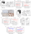
Protein expression of MARCKS and PTEN in IBC and nIBC and prognostic value. (A) Left: protein expression of MARCKS in IBC and nIBC patients (white: negative expression; black: positive expression). Right: forest plots showing the Odds Ratio (log10) of MARCKS expression level in IBC vs. nIBC group in a multivariate logistic regression analysis along with molecular subtypes. (B) Kaplan-Meier MFS curves in nIBC and IBC patients according to MARCKS expression (black: negative; red: positive). (C) Immunohistochemistry staining of PTEN expression in IBC vs. nIBC samples. The illustrated images are represented in; (a) a negative expression, (b) a 20% of staining, (c) 70% of staining, and (d) a 100% of staining: the staining was mainly cytoplasm and some nuclear, and 20× as magnification. (D) Similar to (A), but for PTEN expression. (E) Similar to (B), but for PTEN expression. (F) Box plots of PTEN expression (quick score) according to MARCKS expression group in nIBC (left) and IBC (right) samples, showing the negative correlation in IBC only. (G) Similar to (B) but for the combined expression of MARCKS and PTEN (blue: MARCKS-negative/PTEN-positive; red: no MARCKS-negative/PTEN-positive).
Figure 4
Figure 4
MPS peptide specifically suppressed the activation of MARCKS and regulated PTEN/AKT and MAPK pathways in inflammatory breast cancer cells. MARCKS is involved in various cellular processes, particularly in the cytoskeleton control (by phosphorylation of MARCKS), proliferation, cell motility, and cell survival (by the PI3K/AKT pathway). Exposure of PIP2 on the membrane allows PI3K to phosphorylate it into PIP3, activating AKT-mediated signaling. However, this pathway is regulated by various factors, including PTEN. Our representative figure explains in part the mechanistic role of MPS peptide in IBC cells explaining how MARCKS inhibition subsequently inhibited AKT phosphorylation (represented by an orange cross and down arrows) and, more importantly, the upregulation of PTEN (represented by anascending arrows), leading to apoptosis and the downregulation of the MAPK pathway (represented by an orange cross and down arrows). These found regulations potentially led to the cytoskeleton rearrangement, proliferation, cell motility, tumor stemness inhibition, and apoptosis activation.

References

    1. Robertson F.M., Bondy M., Yang W., Yamauchi H., Wiggins S., Kamrudin S., Krishnamurthy S., Le-Petross H., Bidaut L., Player A.N., et al. Inflammatory Breast Cancer: The Disease, the Biology, the Treatment. CA Cancer J. Clin. 2010;60:351–375. doi: 10.3322/caac.20082. - DOI - PubMed
    1. Boussen H., Bouzaiene H., Ben Hassouna J., Dhiab T., Khomsi F., Benna F., Gamoudi A., Mourali N., Hechiche M., Rahal K., et al. Inflammatory Breast Cancer in Tunisia: Epidemiological and Clinical Trends. Cancer. 2010;116:2730–2735. doi: 10.1002/cncr.25175. - DOI - PubMed
    1. Levine P.H., Veneroso C. The Epidemiology of Inflammatory Breast Cancer. Semin. Oncol. 2008;35:11–16. doi: 10.1053/j.seminoncol.2007.11.018. - DOI - PubMed
    1. Manai M., Finetti P., Mejri N., Athimni S., Birnbaum D., Bertucci F., Rahal K., Gamoudi A., Chaffanet M., Manai M., et al. Inflammatory Breast Cancer in 210 Patients: A Retrospective Study on Epidemiological, Anatomo-Clinical Features and Therapeutic Results. Mol. Clin. Oncol. 2019;10:223–230. doi: 10.3892/mco.2018.1773. - DOI - PMC - PubMed
    1. Dawood S., Merajver S.D., Viens P., Vermeulen P.B., Swain S.M., Buchholz T.A., Dirix L.Y., Levine P.H., Lucci A., Krishnamurthy S., et al. International Expert Panel on Inflammatory Breast Cancer: Consensus Statement for Standardized Diagnosis and Treatment. Ann. Oncol. 2011;22:515–523. doi: 10.1093/annonc/mdq345. - DOI - PMC - PubMed

Publication types

MeSH terms