. 2022 Aug 27;11(9):1674.
doi: 10.3390/antiox11091674.

Mulberry Anthocyanins Ameliorate DSS-Induced Ulcerative Colitis by Improving Intestinal Barrier Function and Modulating Gut Microbiota

Affiliations

Mulberry Anthocyanins Ameliorate DSS-Induced Ulcerative Colitis by Improving Intestinal Barrier Function and Modulating Gut Microbiota

Jianling Mo et al. Antioxidants (Basel). .

Abstract

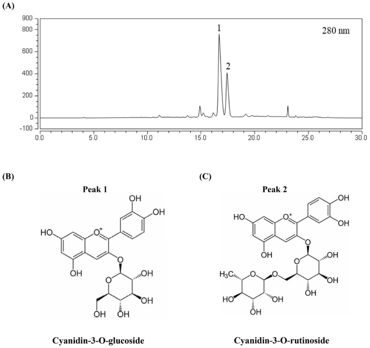
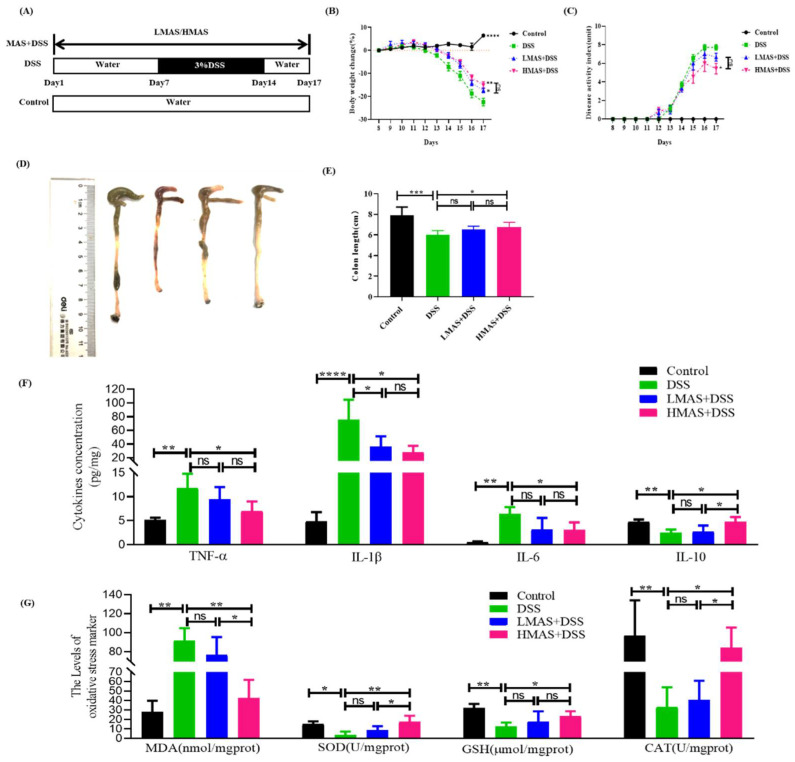
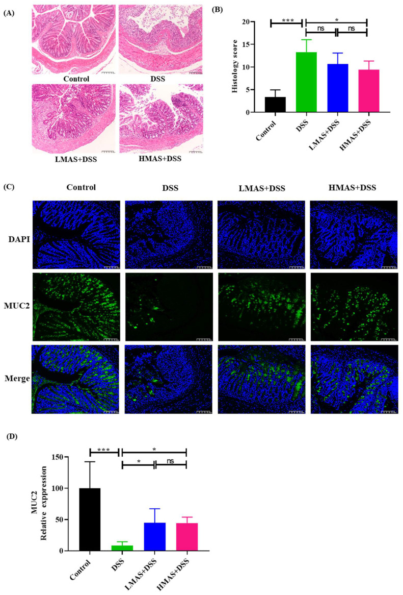
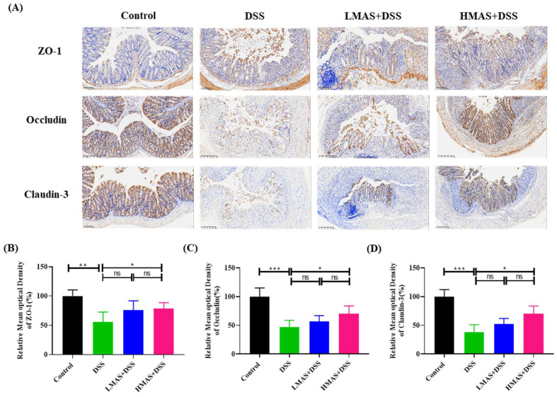
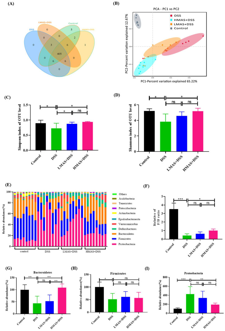
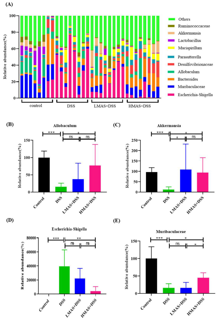
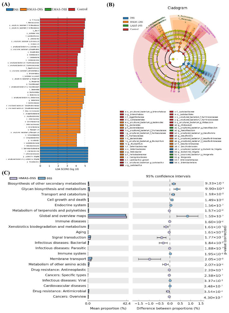
Mulberry has attracted wide attention due to its substantial nutritional values. This work first studied the protective effect of mulberry anthocyanins (MAS) on dextran sulfate sodium (DSS)-induced colitis. The mice experiment was designed as four groups including normal mice (Control), dextran sodium sulfate (DSS)-fed mice, and DSS plus 100 mg/kg·bw MAS-fed mice (LMAS-DSS) or DSS plus 200 mg/kg·bw MAS-fed mice (HMAS-DSS). Mice were given MAS by gavage for 1 week, and then DSS was added to the drinking water for 7 days. MAS was administered for a total of 17 days. The results showed that oral gavage of MAS reduced the disease activity index (DAI), prevented colon shortening, attenuated colon tissue damage and inflammatory response, suppressed colonic oxidative stress and restored the protein expression of intestinal tight junction (TJ) protein (ZO-1, occludin and claudin-3) in mice with DSS-induced colitis. In addition, analysis of 16S rRNA amplicon sequences showed that MAS reduced the DSS-induced intestinal microbiota dysbiosis, including a reduction in Escherichia-Shigella, an increase in Akkermansia, Muribaculaceae and Allobaculum. Collectively, MAS alleviates DSS-induced colitis by maintaining the intestinal barrier, modulating inflammatory cytokines, and improving the microbial community.

Keywords: IBD; gut microbiota; intestinal barrier; mulberry anthocyanins; ulcerative colitis.


Conflict of interest statement

The authors declare that they have no known competing financial interest or personal relationship that could have appeared to influence the work reported in this paper.

Figures

Figure 1
Figure 1
HPLC chromatograms of MAS. (A) The absorbance was detected at 280 nm. (B) Compound name and structure in peak 1. (C) Compound name and structure in peak 2.
Figure 2
Figure 2
MAS attenuates DSS-induced damage. (A) Schematic of experimental design. (B) Changes of body weight. (C) DAI score. (D) Length of experimental mice colon. (E) Representative photograph of colons. (F) The inflammatory TNF-α, IL-1β, IL-6, and anti-inflammatory cytokine IL-10 levels. (G) The levels of oxidative stress markers (MDA, SOD, GSH, and CAT). Here, ns = not significant. Data were expressed as mean ± SD. * p < 0.05, ** p < 0.01, *** p < 0.001, **** p < 0.0001, in comparison with the DSS group (n = 10).
Figure 3
Figure 3
MAS ameliorates intestinal barrier damage in DSS-induced mice. (A) H&E stain of colonic tissues sections (200×). (B) Pathological scores of colonic tissues sections. (C) Muc2 expression in mice colon (200×). The green fluorescence represents the amount of Muc2, and the blue fluorescence is the nucleus stained by DAPI. (D) Quantitative analysis of MUC-2. Here, ns = not significant. Data were expressed as mean ± SD. * p < 0.05, *** p < 0.001, in comparison with the DSS group (n = 10).
Figure 4
Figure 4
MAS reduced intestinal epithelial cell permeability in DSS-induced mice. (A) Immunohistochemistry analysis of ZO-1, Occludin, and Claudin-3; the image magnification is ×200. (BD) Immunohistochemical analysis. Here, ns = not significant. Data are shown as the mean ± SD. * p < 0.05, ** p < 0.01, *** p < 0.001, in comparison with the DSS group (n = 10).
Figure 5
Figure 5
MAS improved the gut microbiota disorder caused by DSS-induced colitis in mice. (A) Venn diagram showing the overlap of the operational taxonomic units (OTUs) identified in the gut microbiota among the four groups. (B) beta diversity analysis of intestinal microbiota among the four groups using the PCA method. (C) Simpson index of all samples. (D) Shannon index of all samples. (E) Bar chart of the bacterial community composition at the phylum level. (H) The Firmicutes to Bacteroidetes ratio. (F,G) Relative abundance of Bacteroidetes, Firmicutes at the phylum level, (I) Proteobacteria at the phylum level. Here, ns = not significant Data are shown as the mean ± SD. * p < 0.05, ** p < 0.01, *** p < 0.001, in comparison with the DSS group (n = 8).
Figure 6
Figure 6
MAS regulates the gut microbiota composition at the genus level. (A) Histogram of the relative abundance of genera among the different groups. (BE) MAS regulated Allobaculum, Akkermansia, Escherichia-Shigella, and Muribaculaceae. Here, ns = not significant. Data are shown as the mean ± SD. * p < 0.05, ** p < 0.01, *** p < 0.001, in comparison with the DSS group (n = 8).
Figure 7
Figure 7
MAS regulates the bacterial metabolic pathway. (A) LDA analysis. (B) LEfSe analysis. (C) Analysis of differences in KEGG metabolic pathways between groups.

References

    1. Ungaro R., Mehandru S., Allen P.B., Peyrin-Biroulet L., Colombel J.F. Ulcerative colitis. Lancet. 2017;389:1756–1770. doi: 10.1016/S0140-6736(16)32126-2. - DOI - PMC - PubMed
    1. Torres J., Mehandru S., Colombel J.F., Peyrin-Biroulet L. Crohn’s disease. Lancet. 2017;389:1741–1755. doi: 10.1016/S0140-6736(16)31711-1. - DOI - PubMed
    1. Kobayashi T., Siegmund B., Le Berre C., Wei S.C., Ferrante M., Shen B., Bernstein C.N., Danese S., Peyrin-Biroulet L., Hibi T. Ulcerative colitis. Nat. Rev. Dis. Primers. 2020;6:74. doi: 10.1038/s41572-020-0205-x. - DOI - PubMed
    1. Annese V. Genetics and epigenetics of IBD. Pharmacol. Res. 2020;159:104892. doi: 10.1016/j.phrs.2020.104892. - DOI - PubMed
    1. Kudelka M.R., Stowell S.R., Cummings R.D., Neish A.S. Intestinal epithelial glycosylation in homeostasis and gut microbiota interactions in IBD. Nat. Rev. Gastroenterol. Hepatol. 2020;17:597–617. doi: 10.1038/s41575-020-0331-7. - DOI - PMC - PubMed

LinkOut - more resources