. 2022 Aug 30;11(9):1702.
doi: 10.3390/antiox11091702.

Intrarenal Arterial Transplantation of Dexmedetomidine Preconditioning Adipose Stem-Cell-Derived Microvesicles Confers Further Therapeutic Potential to Attenuate Renal Ischemia/Reperfusion Injury through miR-122-5p/Erythropoietin/Apoptosis Axis

Affiliations

Intrarenal Arterial Transplantation of Dexmedetomidine Preconditioning Adipose Stem-Cell-Derived Microvesicles Confers Further Therapeutic Potential to Attenuate Renal Ischemia/Reperfusion Injury through miR-122-5p/Erythropoietin/Apoptosis Axis

Yu-Hsuan Cheng et al. Antioxidants (Basel). .

Abstract

Intravenous adipose mesenchymal stem cells (ADSCs) attenuate renal ischemia/reperfusion (IR) injury but with major drawbacks, including the lack of a specific homing effect after systemic infusion, cell trapping in the lung, and early cell death in the damaged microenvironment. We examined whether intrarenal arterial transplantation of dexmedetomidine (DEX) preconditioning ADSC-derived microvesicles (DEX-MVs) could promote further therapeutic potential to reduce renal IR injury. We evaluated the effect of DEX-MVs on NRK-52E cells migration, hypoxia/reoxygenation (H/R)-induced cell death, and reactive oxygen species (ROS) amount and renal IR model in rats. IR was established by bilateral 45 min ischemia followed by 4 h reperfusion. Intrarenal MVs or DEX-MVs were administered prior to ischemia. Renal oxidative stress, hemodynamics and function, western blot, immunohistochemistry, and tubular injury scores were determined. The miR-122-5p expression in kidneys was analyzed using microarrays and quantitative RT-PCR and its action target was predicted by TargetScan. DEX-MVs were more efficient than MVs to increase migration capability and to further decrease H/R-induced cell death and ROS level in NRK-52E cells. Consistently, DEX-MVs were better than MV in increasing CD44 expression, improving IR-depressed renal hemodynamics and renal erythropoietin expression, inhibiting IR-enhanced renal ROS level, tubular injury score, miR-122-5p expression, pNF-κB expression, Bax/caspase 3/poly(ADP-ribose) polymerase (PARP)-mediated apoptosis, blood urea nitrogen, and creatinine levels. The use of NRK-52E cells confirmed that miR-122-5p mimic via inhibiting erythropoietin expression exacerbated Bax-mediated apoptosis, whereas miR-122-5p inhibitor via upregulating erythropoietin and Bcl-2 expression reduced apoptosis. In summary, intrarenal arterial DEX-MV conferred further therapeutic potential to reduce renal IR injury through the miR-122-5p/erythropoietin/apoptosis axis.

Keywords: adipose-derived mesenchymal stem cells; apoptosis; dexmedetomidine preconditioning; erythropoietin; ischemia/reperfusion; miR-122-5p; microvesicles.


Conflict of interest statement

The authors declare no conflict of interest.

Figures

Figure 1
Figure 1
Identification of purified ADSC with CD90-positive characterization and CD45-negative characterization in the P4 ADSC by flow cytometry (A). The morphology in control ADSC (ADSC) and DEX-treated ADSC (DEX-ADSC) (Magnification Scale: 50 μm) in 80% confluency is similar in our result (B). Cells displayed fibroblast-like morphology (long and thin) under phase-contrast microscope. Nanoparticle size of MV is amplified 50,000 times (left) and 100,000 times (right), existing in ADSC culture medium (C). Effect of MVs and DEX-MVs on wound healing of NRK-52E cells in different time courses (D). DEX-MVs significantly decreased wound size ratio as compared to MVs. NRK-52E cells in response to H/R significantly decreased viability (E) and increased ROS (F) vs. CON. MVs or DEX-MVs significantly improved cell viability and reduced ROS counts vs. H/R group. DEX-MVs were more efficient than MVs in increase in cell viability and decrease in ROS counts. n = 6 in each experiment. ADSC, adipose stem cell; DEX, dexmedetomidine; MV, microvesicle; H/R, hypoxia/reoxygenation; ROS, reactive oxygen species. * p < 0.05 vs. CON. # p < 0.05 vs. H/R. a p < 0.05 vs. MV.
Figure 2
Figure 2
(A): The experimental protocol for intrarenal arterial injection of fluorescent MV to the kidney. (B): Detection of fluorescence expression in kidneys of rats treated with ADSC-derived MVs (n = 6). Representative fluorescent micrographs showing the expression of CD44 proteins in kidney sections of four groups of rats and sacrificed 1 h later. Original magnification: ×400. Arrows indicate fluorescent MVs. Six animals per groups were examined with similar results. (C): Western blot analysis of CD 44 expression of kidney homogenates in four groups of kidneys. (D): Statistical data of CD44 expression by Western blot (n = 3 each). (E): Statistical data of fluorescent MV expression of four groups of kidneys (n = 6 each). ADSC, adipose stem cell; MV, microvesicle; IR, ischemia/reperfusion; IRMV, ischemia/reperfusion with microvesicle. * p < 0.05 vs. CON rat. # p < 0.05 vs. IR rat. a p < 0.05 vs. IRMV rat.
Figure 3
Figure 3
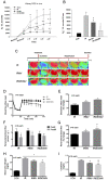
In response to renal IR injury, in vivo kidney ROS measurement was indicated in four groups of rats (n = 6 each) (A). The average ROS amount (n = 6 each) was displayed in (B). Renal microcirculation was determined with a moor image in three IR groups of rats (C). Renal IR significantly decreased renal microcirculation, as indicated by PU value in IR rats vs. IRMV and IRDEXMV treated rats (n = 6 each) (D). Renal PU significantly recovered towards the normal value within 10 min reperfusion in IRMV- and-IRDEXMV treated rats (n = 6 each). (E): The percentage change of blood flow influx mean is shown. Renal arterial blood flow is significantly decreased in IR and IRMV rats, but not in IRDEXMV rats (n = 6 each) (F). Percentage change of renal arterial blood flow is significantly increased in IRMV− and IRDEXMV−treated rats vs. IR rats within 10 min reperfusion (n = 6 each) (G). Renal IR significantly increases blood urea nitrogen (H) and creatinine (I) levels in IR−, IRMV−, and IRDEXMV− treated rats (n = 6 each). However, these two levels are significantly decreased in IRDEXMV vs. IR rats. * p < 0.05 vs. CON rat. MV, microvesicle; IR, ischemia/reperfusion; IRMV, ischemia/reperfusion with microvesicle; IRDEXMR, ischemia/reperfusion with dexmedetomidine preconditioned microvesicle; PU, perfusion unit; ROS, reactive oxygen species. # p < 0.05 vs. IR rat. a p < 0.05 vs. IRMV rat.
Figure 4
Figure 4
Effect of MVs or DEXMVs on pathologic parameters in renal cortex and medulla by H&E stain (A), pNF-κB, Caspase-3, and PARP stain (B) in CON, IR, MV, and DEXMV groups. The statistical data of tubular injury score (C), p-NF-κB expression (D), Caspase-3 expression, (E) and PARP expression (F) are indicated. Data are expressed as mean ± SEM in each group (n = 6) using single values. * p < 0.05 compared with CON group. td: tubular dilation. Yellow arrows in (A) indicate tubular dilation (td). Asterisks in (A) indicate erythrocyte accumulation. Yellows in (B) indicate each brown stain. MV, microvesicle; IR, ischemia/reperfusion; IRMV, ischemia/reperfusion with microvesicle; IRDEXMR, ischemia/reperfusion with dexmedetomidine preconditioned microvesicle; PARP, poly-(ADP-ribose)-polymerase; SEM, standard error mean. # p < 0.05 compared with IR group. a p < 0.05 vs. MV group.
Figure 5
Figure 5
miRNA expression in four groups of kidneys. (A) The heat map shows the expression level of whole−kidney miRNAs in CON (S1, S2, S3), IR (IR1, IR2, IR3), IRMV (IRMV1, IRMV2, IRMV3), and IRDEXMV (DEXMV1, DEXMV2, DEXMV3) (n = 3 each). Each column represents a sample and each row represents a miRNA in the graph. The expression ratio is represented by color ranges from green (low) to red (high), as indicated by the scale bar. (B) The highest relative expression level of miR-122-5p in microarray is analyzed and displayed in these four groups. (C) Relative expression of miR-122-5p in kidney is detected by quantitative RT−PCR. (D) To understand the mechanism whereby miR-122-5p contributes to IR, by using online databases (TargetScan), we have identified a conserved putative miR-122-5p-targeting site in the EPO mRNA. (E) Erythropoietin (EPO) mRNA levels from four groups of kidneys were determined by quantitative RT−PCR. Relative fold-change values were normalized against GAPDH as endogenous control and expressed as fold changes over sham control kidney. (F) Luciferase activity of EPO in four groups of kidneys. Data are expressed as mean ± SEM in each group (n = 3) using single values. * p < 0.05 compared with CON group. # p < 0.05 compared with IR group. EPO, erythropoietin; MV, microvesicle; IR, ischemia/reperfusion; IRMV, ischemia/reperfusion with microvesicle; IRDEXMR, ischemia/reperfusion with dexmedetomidine preconditioned microvesicle; RT−PCR, real−time polymerase chain reaction; SEM, standard error mean. a p < 0.05 vs. IRMV group.
Figure 6
Figure 6
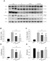
Effect of ADSC-derived MVs (IRMV) or DEX preconditioning ADSC-derived MVs (IRDEXMV) on Bax, Bcl-2, cleaved caspase-3 (c-caspase-3), PARP, and EPO expression in the four groups of kidneys. (A): The original data of Western blot. (B): Statistical data of the ratio of Bax/Bcl-2. (C): Statistical data of the ratio of C-Casp 3/actin. (D): Statistical data of the ratio of PARP/actin. (E): Statistical data of the ratio of EPO/actin. Data are expressed as mean ± SEM in each group (n = 3) using the single values. * p < 0.05 compared with CON group. ADSC, adipose stem cells; c-caspase-3, cleaved caspase-3; EPO, erythropoietin; MV, microvesicle; IR, ischemia/reperfusion; IRMV, ischemia/reperfusion with microvesicle; IRDEXMR, ischemia/reperfusion with dexmedetomidine preconditioned microvesicle; PARP, poly-(ADP-ribose)-polymerase; SEM: standard error mean. # p < 0.05 compared with IR group. a p < 0.05 compared with IR+MV group.
Figure 7
Figure 7
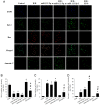
Effect of miR-122-5p mimics and inhibitor on H/R-induced Bcl-2, Bax, and apoptosis formation in the NRK-52E cells. (A): The immunofluorescence graphs of DAPI (blue), Bcl-2 (green), Bax (red), merged image, and Annexin V (green) in response to Scheme 2 positive cell density; (B): Bcl-2 positive cell density, (C): Bax positive cell density and (D): Annexin V positive cell density. Data are expressed as mean ± SEM (n = 3). * p < 0.05 compared with CON group. DAPI, 4’, 6-diamidino-2-phenylindole; EPO, erythropoietin; H/R, hypoxia/reoxygenation; SEM, standard error mean. # p < 0.05 compared with H/R group. a p < 0.05 vs. H/R+miR-122-5p m group.
Figure 8
Figure 8
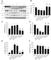
Effect of H/R, miR-122-5p mimics (m), miR-122-5p inhibitor (I), or EPO on EPO, Bax, Bcl-2, C-Casp 3, and PARP expression in NRK-52E cells by Western blot (A). (B) Statistic data of EPO expression; (C) statistic data of Bax expression; (D) statistic data of Bcl-2 expression; (E) statistic data of C-Caspase 3 expression; and (F) Statistic data of PARP expression. Data are expressed as mean ± SEM (n = 3). C-Casp 3, cleaved caspase 3; EPO, erythropoietin; H/R, hypoxia/reoxygenation; PARP, poly-(ADP-ribose)-polymerase; SEM: standard error mean. * p < 0.05 compared with CON group. # p < 0.05 compared with H/R group.
Figure 9
Figure 9
The summary diagram. The summary diagram suggests that DEX-preconditioned ADSC-derived MVs can provide CD44 homing effect; ameliorate IR-induced renal hemodynamics depression; and reduce renal ROS level, tubular cell inflammation, apoptosis, tubular injury score, and renal dysfunction through downregulated miR-122-5p/Bax/PARP/apoptosis signaling and upregulated target-gene EPO expression in the post-IR kidney. DEX-preconditioned ADSC-derived MVs could be an important tool for utilization during renal IR injury. ADSC, adipose stem cells; AKI, acute kidney injury; DEX, dexmedetomidine; EPO, erythropoietin; MV, microvesicle; I/R, ischemia/reperfusion; PARP, poly-(ADP-ribose)-polymerase; ROS, reactive oxygen species.

References

    1. Chien C.-T., Chang T.-C., Tsai C.-Y., Shyue S.-K., Lai M.-K. Adenovirus-Mediated bcl-2 Gene Transfer Inhibits Renal Ischemia/Reperfusion Induced Tubular Oxidative Stress and Apoptosis. Am. J. Transplant. 2005;5:1194–1203. doi: 10.1111/j.1600-6143.2005.00826.x. - DOI - PubMed
    1. Yang C.-C., Yao C.-A., Yang J.-C., Chien C.-T. Sialic Acid Rescues Repurified Lipopolysaccharide-Induced Acute Renal Failure via Inhibiting TLR4/PKC/gp91-Mediated Endoplasmic Reticulum Stress, Apoptosis, Autophagy, and Pyroptosis Signaling. Toxicol. Sci. 2014;141:155–165. doi: 10.1093/toxsci/kfu121. - DOI - PMC - PubMed
    1. Kim D.-W., Staples M., Shinozuka K., Pantcheva P., Kang S.-D., Borlongan C.V. Wharton’s Jelly-Derived Mesenchymal Stem Cells: Phenotypic Characterization and Optimizing Their Therapeutic Potential for Clinical Applications. Int. J. Mol. Sci. 2013;14:11692–11712. doi: 10.3390/ijms140611692. - DOI - PMC - PubMed
    1. Deng Z.-B., Poliakov A., Hardy R.W., Clements R., Liu C., Liu Y., Wang J., Xiang X., Zhang S., Zhuang X., et al. Adipose Tissue Exosome-Like Vesicles Mediate Activation of Macrophage-Induced Insulin Resistance. Diabetes. 2009;58:2498–2505. doi: 10.2337/db09-0216. - DOI - PMC - PubMed
    1. Altadill T., Campoy I., Lanau L., Gill K., Rigau M., Gil-Moreno A., Reventos J., Byers S., Colas E., Cheema A.K. Enabling Metabolomics Based Biomarker Discovery Studies Using Molecular Phenotyping of Exosome-Like Vesicles. PLoS ONE. 2016;11:e0151339. doi: 10.1371/journal.pone.0151339. - DOI - PMC - PubMed