

Homocysteine and Inflammatory Cytokines in the Clinical Assessment of Infection in Venous Leg Ulcers
- PMID: 36140047
- PMCID: PMC9495878
- DOI: 10.3390/antibiotics11091268
Homocysteine and Inflammatory Cytokines in the Clinical Assessment of Infection in Venous Leg Ulcers
Abstract
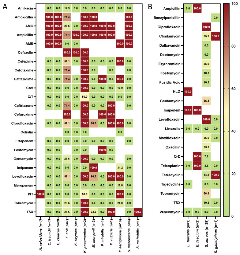
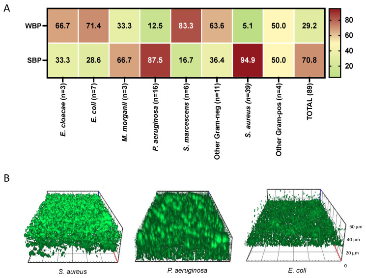
Inflammation and biofilm-associated infection are common in chronic venous leg ulcers (VU), causing deep pain and delayed healing. Albeit important, clinical markers and laboratory parameters for identifying and monitoring persistent VU infections are limited. This study analyzed 101 patients with infected (IVU) and noninfected VUs (NVU). Clinical data were collected in both groups. The serum homocysteine (Hcys) and inflammatory cytokines from the wound fluid were measured. In addition, microbial identification, antibiotic susceptibility, and biofilm production were examined. IVU were 56 (55.4%) while NVU were 45 (44.5%). IVUs showed a significant increase in the wound's size and depth compared to NVUs. In addition, significantly higher levels of interleukin (IL)-6, IL-10, IL17A, and tumor necrosis factor-alpha (TNF-α) were found in patients with IVUs compared to those with NVUs. Notably, hyperhomocysteinemia (HHcy) was significantly more common in patients with IVUs than NVUs. A total of 89 different pathogens were identified from 56 IVUs. Gram-negative bacteria were 51.7%, while the Gram-positives were 48.3%. At the species level, Staphylococcus aureus was the most common isolate (43.8%), followed by Pseudomonas aeruginosa (18.0%). Multidrug-resistant organisms (MDROs) accounted for 25.8% of the total isolates. Strong biofilm producers (SBPs) (70.8%) were significantly more abundant than weak biofilm producers (WBP) (29.2%) in IVUs. SBPs were present in 97.7% of the IVUs as single or multispecies infections. Specifically, SBPs were 94.9% for S. aureus, 87.5% for P. aeruginosa, and 28.6% for Escherichia coli. In IVU, the tissue microenvironment and biofilm production can support chronic microbial persistence and a most severe clinical outcome even in the presence of an intense immune response, as shown by the high levels of inflammatory molecules. The measurement of local cytokines in combination with systemic homocysteine may offer a novel set of biomarkers for the clinical assessment of IVUs caused by biofilm-producing bacteria.
Keywords: biofilm; cytokines; homocysteine; infection; leg ulcers; venous ulcer; wound.
Conflict of interest statement
The authors declare no conflict of interest.
Figures
References
LinkOut - more resources
Full Text Sources
Molecular Biology Databases
