. 2022 Aug 27;12(9):691.
doi: 10.3390/bios12090691.

Use of Deep Learning to Detect the Maternal Heart Rate and False Signals on Fetal Heart Rate Recordings

Affiliations

Use of Deep Learning to Detect the Maternal Heart Rate and False Signals on Fetal Heart Rate Recordings

Samuel Boudet et al. Biosensors (Basel). .

Abstract

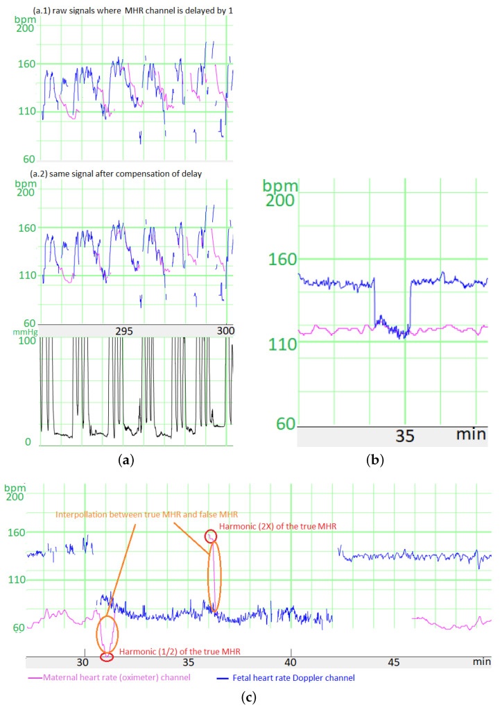
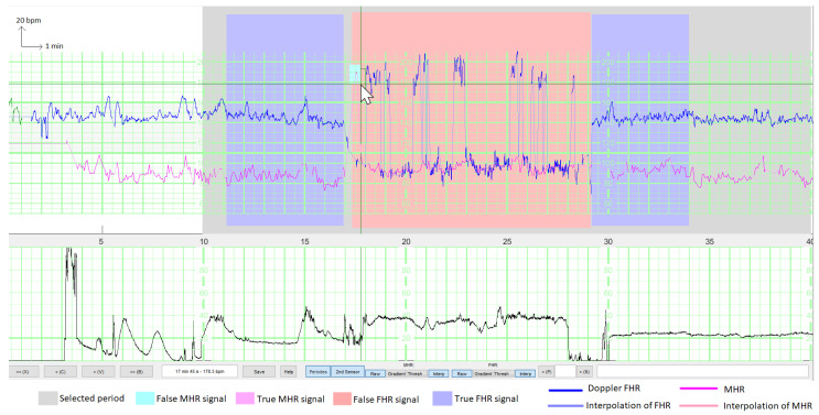
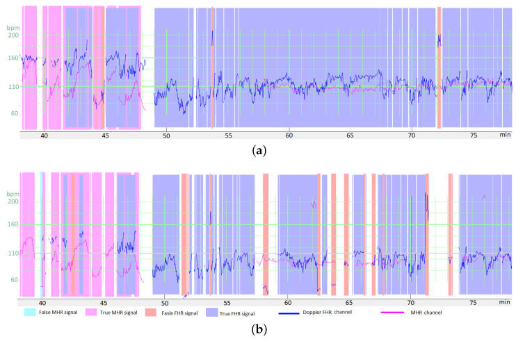
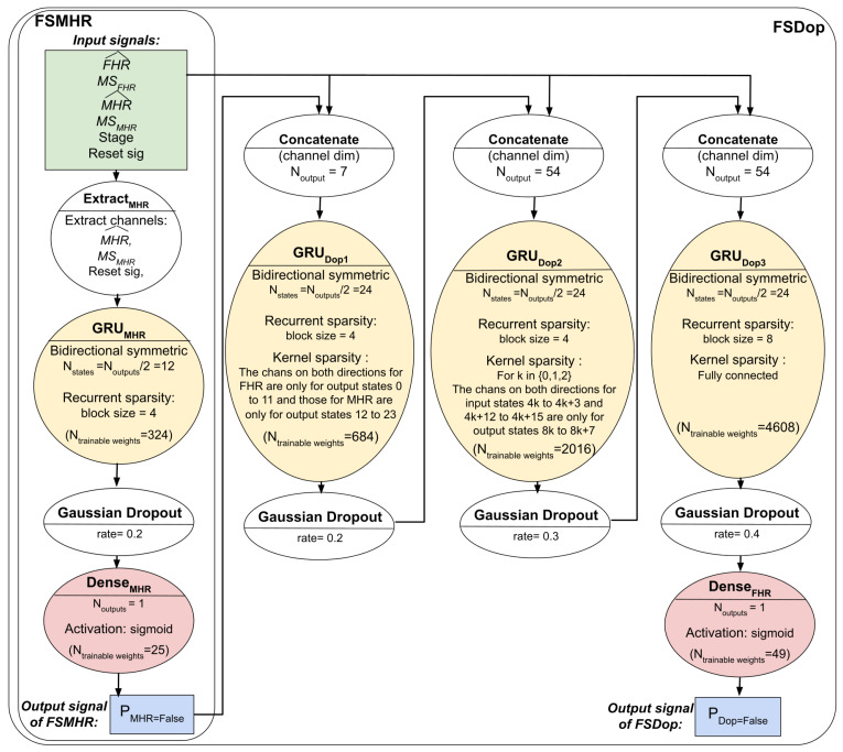
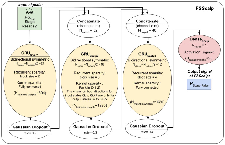
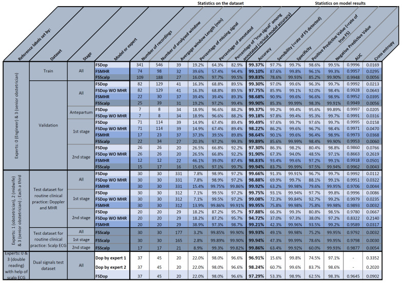
We have developed deep learning models for automatic identification of the maternal heart rate (MHR) and, more generally, false signals (FSs) on fetal heart rate (FHR) recordings. The models can be used to preprocess FHR data prior to automated analysis or as a clinical alert system to assist the practitioner. Three models were developed and used to detect (i) FSs on the MHR channel (the FSMHR model), (ii) the MHR and FSs on the Doppler FHR sensor (the FSDop model), and (iii) FSs on the scalp ECG channel (the FSScalp model). The FSDop model was the most useful because FSs are far more frequent on the Doppler FHR channel. All three models were based on a multilayer, symmetric, GRU, and were trained on data recorded during the first and second stages of delivery. The FSMHR and FSDop models were also trained on antepartum recordings. The training dataset contained 1030 expert-annotated periods (mean duration: 36 min) from 635 recordings. In an initial evaluation of routine clinical practice, 30 fully annotated recordings for each sensor type (mean duration: 5 h for MHR and Doppler sensors, and 3 h for the scalp ECG sensor) were analyzed. The sensitivity, positive predictive value (PPV) and accuracy were respectively 62.20%, 87.1% and 99.90% for the FSMHR model, 93.1%, 95.6% and 99.68% for the FSDop model, and 44.6%, 87.2% and 99.93% for the FSScalp model. We built a second test dataset with a more solid ground truth by selecting 45 periods (lasting 20 min, on average) on which the Doppler FHR and scalp ECG signals were recorded simultaneously. Using scalp ECG data, the experts estimated the true FHR value more reliably and thus annotated the Doppler FHR channel more precisely. The models achieved a sensitivity of 53.3%, a PPV of 62.4%, and an accuracy of 97.29%. In comparison, two experts (blinded to the scalp ECG data) respectively achieved a sensitivity of 15.7%, a PPV of 74.3%, and an accuracy of 96.91% and a sensitivity of 60.7%, a PPV of 83.5% and an accuracy of 98.24%. Hence, the models performed at expert level (better than one expert and worse than the other), although a well-trained expert with good knowledge of FSs could probably do better in some cases. The models and datasets have been included in the Fetal Heart Rate Morphological Analysis open-source MATLAB toolbox and can be used freely for research purposes.

Keywords: cardiotocogram; deep learning; fetal heart rate; gated recurrent unit; maternal heart rate.


Conflict of interest statement

The authors have no conflict of interest to declare.

Figures

Figure A1
Figure A1
Illustration of the effects produced by CTG monitors, complicating the recognition of FSs. These effects are due to the autocorrelation algorithm used to estimate heart rate. (a) Example of an FHR FS in a second stage recording, with and without correction for the lag in the MHR channel. (b) Example of a continuous transition over ≈1 s in the FHR signal. (c) Example of a continuous transition over ≈20 s in the MHR signal measured with finger oximeter, when switching to an FS. This was due to the broad autocorrelation window used by the CTG monitor to compute the MHR from the oximeter signal.
Figure 1
Figure 1
Illustration of the interface for annotating signals during a period with an epidural injection. Both the FHR (in blue) and the MHR (in purple) are shown, together with their interpolations (lighter colors) during MS periods (for better visualization of samples that are isolated from the rest of the signal). The user has selected a window for training the model (in grey) and has annotated two periods with a true FHR signal (in blue) and one period with a false FHR signal (in red). The false signals are either the MHR or the MHR × 2. Here, the user is selecting a few false MHR signals (in cyan) inside the rectangular box.
Figure 2
Figure 2
The input matrix for a unit in the FSDop model. FHR^ and MHR^ are the normalized FHR and the normalized MHR, respectively; MSFHR and MSMHR are respectively binary channels indicating whether the heart rate is an MS; Stage is a binary channel indicating whether the sample is in the first or second stage of delivery.
Figure 3
Figure 3
Example of the transformation of a period for data augmentation, with relatively high changes in the Doppler FHR channel (in blue) and the MHR channel (in purple). (a) The raw data in a period. (b) The period after random transformation. Expert annotations are shown as colored zones. Data augmentation consisted in adding MS and FS on both the FHR and MHR channels. Moreover, both the MHR and the FHR were multiplied by a λ0.9. The final signal corresponds to poor quality recordings but remains realistic. It should be noted that the MHR and FHR were not annotated by the experts for the entire recording (either because the experts were uncertain, or the period did not contain difficult-to-interpret features).
Figure 4
Figure 4
The FSMHR and FSDop models’ architectures and hyperparameters. For each MHR signal sample, FSMHR outputs the probability of being an FS and for each Doppler FHR signal sample, FSDop outputs the probability of being an FS.
Figure 5
Figure 5
The FSScalp model’s architectures and hyperparameters. For each Scalp ECG FHR signal sample, FSScalp outputs the probability of being an FS.
Figure 6
Figure 6
Examples of results for the three models (FSMHR, FSDop and FSScalp). (a) Results for FSScalp with a recording from the first stage of delivery. (b) Results for FSScalp with a recording from the first stage of delivery. (c) Results for FSDop and FSMHR with a recording from the first stage of delivery. (d) Results for FSDop and FSMHR with a recording from the first stage of delivery, during the administration of an epidural. The likelihood of an FS estimated by each model is represented as a color gradient. On examples (a,c,d), the blue/red signal corresponds to the Doppler channel and the green signal corresponds to the scalp ECG channel. On example (b), the blue/red signal is the scalp ECG signal. On all recordings, the time lag in the MHR had been corrected (as described in Section 2.4).
Figure 7
Figure 7
The FHRMA toolbox interface automatically displays both FSs and the results of a morphological analysis (baseline, accelerations, decelerations, and UCs). The FHR signal comes from the Doppler sensor. The second stage of delivery starts at 475 min.
Figure 8
Figure 8
Statistical results for the study’s models and datasets.

References

    1. Ayres-de-Campos D., Spong C.Y., Chandraharan E. FIGO Consensus Guidelines on Intrapartum Fetal Monitoring: Cardiotocography. Int. J. Gynecol. Obstet. 2015;131:13–24. doi: 10.1016/j.ijgo.2015.06.020. - DOI - PubMed
    1. Health Encyclopedia. University of Rochester Medical Center; Rochester, NY, USA: [(accessed on 19 July 2022)]. External and Internal Heart Rate Monitoring of the Fetus. Available online: https://www.urmc.rochester.edu/encyclopedia/content.aspx?contenttypeid=9....
    1. Maternia A., Kupka T., Horoba K., Jezewski J., Martinek R., Wrobel J., Kahankova R., Czabanski R., Graczyk S. New Possibilities for Fetal Monitoring Using Unobtrusive Abdominal Electrocardiography; Proceedings of the 2019 MIXDES—26th International Conference “Mixed Design of Integrated Circuits and Systems”; Rzeszów, Poland. 27–29 June 2019; pp. 413–418. - DOI
    1. Lee K.J., Lee B. End-to-End Deep Learning Architecture for Separating Maternal and Fetal ECGs Using W-Net. IEEE Access. 2022;10:39782–39788. doi: 10.1109/ACCESS.2022.3166925. - DOI
    1. Odendaal H.J. False Interpretation of Fetal Heart Role Monitoring in Cases of Intra-Uterine Death. S. Afr. Med. J. 1976;50:1963–1965. - PubMed