Review
. 2022 Sep 6;12(9):731.
doi: 10.3390/bios12090731.

Carbon Nanotube and Its Derived Nanomaterials Based High Performance Biosensing Platform

Affiliations
Review

Carbon Nanotube and Its Derived Nanomaterials Based High Performance Biosensing Platform

Jagannath Mondal et al. Biosensors (Basel). .

Abstract

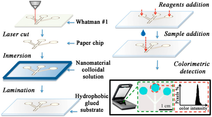
After the COVID-19 pandemic, the development of an accurate diagnosis and monitoring of diseases became a more important issue. In order to fabricate high-performance and sensitive biosensors, many researchers and scientists have used many kinds of nanomaterials such as metal nanoparticles (NPs), metal oxide NPs, quantum dots (QDs), and carbon nanomaterials including graphene and carbon nanotubes (CNTs). Among them, CNTs have been considered important biosensing channel candidates due to their excellent physical properties such as high electrical conductivity, strong mechanical properties, plasmonic properties, and so on. Thus, in this review, CNT-based biosensing systems are introduced and various sensing approaches such as electrochemical, optical, and electrical methods are reported. Moreover, such biosensing platforms showed excellent sensitivity and high selectivity against not only viruses but also virus DNA structures. So, based on the amazing potential of CNTs-based biosensing systems, healthcare and public health can be significantly improved.

Keywords: carbon nanotubes; high-performance biosensors; nanomaterials-based biosensors.


Conflict of interest statement

The authors declare no conflict of interest.

Figures

Figure 1
Figure 1
Schematic presentation of CNT-based biosensors.
Figure 2
Figure 2
Biodistribution of Gallium-67-labeled single-walled carbon nanotubes in murine breast cancer model. (A) SPECT/CT images of tumor-bearing mice after 24 h of injection of either Ga-SWCNT or Ga-SWCNT + CD44 (right). (B) Quantitative measurement of radioactivity of Ga-SWCNT or Ga-SWCNT + CD44 at various organs and tumor after dosing at different time points: 2, 24, and 48 h represented as dose per gram of tissue. Image reproduced with permission from [134]. Copyright 2016 Future Medicine Ltd.
Figure 3
Figure 3
(a) Functionalization and fabrication of CNTs chip. In order to fabricate the chip CNT film was allowed to adsorb on the glass substrate followed by sealing with PDMS cover. Biotin-PLL was attached to the CNT film via Pi and hydrophobic interaction. (b) Schematic illustration showing the release mechanism of captured CTCs. There will be deprotonation of positively charged amino groups causing the conformational changes in the PLL structure of random coil to alfa-helix resulting in cancellation of interaction between PLL and CNTs releasing the captured CTCs. Image reproduced with permission from [135]. Copyright 2022 American Chemical Society.
Figure 4
Figure 4
Using CNTs and PEI layer-by-layer (LbL) assembly was constructed on gold surface to fabricate the thin film. Carboxylic acid groups on the CNT surface were activated using EDC-NHS reagents to attach the anti-CA19-9 antibodies. Right side image shows the antibody–antigen reaction. Image reproduced with permission from [137]. Copyright 2017 American Chemical Society.
Figure 5
Figure 5
Schematic illustration showing the fabrication of nano-sensor device: preparation of nanocomposite of PANI/CNT and assembly of aptamer VEGF on the sensor surface. Image reproduced with permission from [139].
Figure 6
Figure 6
(A) Schematic illustration of the nanoprobe fabrication steps. (B) Working electrode modification along with immunosensor principle based on direct competitive electrochemical sensor for MUC−1 detection. Several steps are involved: (1) Electro oxidative grafting of gelatin on electrode; (2) MUC−1 antibody binding with EDC/NHS; (3) attaching nanoprobe with modified electrode; (4) decrease in signal after free MUC−1 replaced nanoprobe in a competitive assay. Image reproduced with permission from [140]. Copyright 2020 Elsevier.
Figure 7
Figure 7
(i, ii) Nanohybrid preparation: SWCNT was modified with reduced graphene oxide (which was prepared by Improved Hummers Method) and cobalt phthalocyanine. The SEM image corresponds to SWCNT/rGO/CoPc nanohybrid. (iii) Fabrication of sensing electrodes (glassy carbon electrode or GCE) by drop-casting method. (iv) Electrochemical oxidation of D-glucose to gluconic acid. (a) Static chronoamperogram response of GCE-SWCNT/rGO/CoPc with increasing glucose concentrations from 0 mM to 5.0 mM in 0.10 M NaOH, and (b) the corresponding calibration plot of steady-state current against concentrations of glucose.. Image reproduced with permission from [182]. Copyright 2021 Elsevier.
Figure 8
Figure 8
Schematic illustration of disposable microfluidic paper-based devices (μPADs). Image reproduced with permission from [183]. Copyright 2015 American Chemical Society.
Figure 9
Figure 9
Schematic illustration of the functionalization of CNT and enzyme immobilization method. Image reproduced with permission from [169]. Copyright 2021 Elsevier.
Figure 10
Figure 10
Schematic illustration of GF/CNTs/GNPs preparation for uric acid detection biosensor. Image reproduced with permission from [207]. Copyright 2017 Elsevier.
Figure 11
Figure 11
Schematic representation of fabrication of ACE2-(GT)6-SWCNTs nanosensor using ACE2 as a sensing protein. Image reproduced with permission from [179]. Copyright 2021 American Chemical Society.
Figure 12
Figure 12
Schematic illustration of SARS-CoV-2 S1 testing steps of CNT-FET biosensor. Anti-SARS-CoV-2 S1 was conjugated on the CNT (using PBASE as a linker) to produce SARS-CoV-2 S1 detectable CNT-FET biosensor. Image reproduced with permission from [28]. Copyright 2021 Elsevier.
Figure 13
Figure 13
Schematic illustration of fabrication of magnetically aligned gold/magnetic nanoparticles decorated CNT on Pt-IDE for virus DNA-sensing platform. Image reproduced with permission from [219]. Copyright 2018 Elsevier.

Similar articles

Cited by

References

    1. Justino C.I.L., Rocha-Santos T.A., Duarte A.C., Rocha-Santos T.A. Review of Analytical Figures of Merit of Sensors and Biosensors in Clinical Applications. TrAC Trends Anal. Chem. 2010;29:1172–1183. doi: 10.1016/j.trac.2010.07.008. - DOI
    1. Guo S., Dong S. Biomolecule-Nanoparticle Hybrids for Electrochemical Biosensors. TrAC Trends Anal. Chem. 2009;28:96–109. doi: 10.1016/j.trac.2008.10.014. - DOI
    1. Chen X., Van Pée K.-H. Catalytic Mechanisms, Basic Roles, and Biotechnological and Environmental Significance of Halogenating Enzymes. Acta Biochim. Biophys. Sin. 2008;40:183–193. doi: 10.1111/j.1745-7270.2008.00390.x. - DOI - PubMed
    1. Cornell B.A., Braach-Maksvytis V.L.B., King L.G., Osman P.D.J., Raguse B., Wieczorek L., Pace R.J. A Biosensor That Uses Ion-Channel Switches. Nature. 1997;387:580–583. doi: 10.1038/42432. - DOI - PubMed
    1. Luong J.H.T., Male K.B., Glennon J.D. Biosensor Technology: Technology Push versus Market Pull. Biotechnol. Adv. 2008;26:492–500. doi: 10.1016/j.biotechadv.2008.05.007. - DOI - PubMed