. 2022 Aug 29;10(9):2117.
doi: 10.3390/biomedicines10092117.

The Gelatinase Inhibitor ACT-03 Reduces Gliosis in the Rapid Kindling Rat Model of Epilepsy, and Attenuates Inflammation and Loss of Barrier Integrity In Vitro

Affiliations

The Gelatinase Inhibitor ACT-03 Reduces Gliosis in the Rapid Kindling Rat Model of Epilepsy, and Attenuates Inflammation and Loss of Barrier Integrity In Vitro

Diede W M Broekaart et al. Biomedicines. .

Abstract

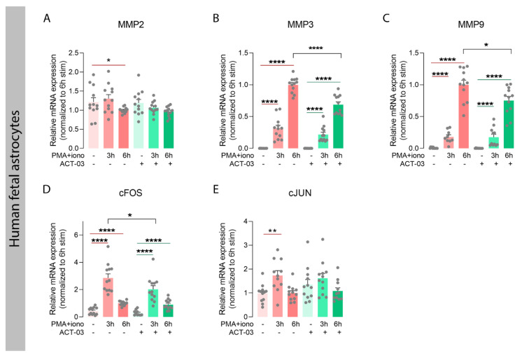
Matrix metalloproteinases (MMPs) are endopeptidases responsible for the cleavage of intra- and extracellular proteins. Several brain MMPs have been implicated in neurological disorders including epilepsy. We recently showed that the novel gelatinase inhibitor ACT-03 has disease-modifying effects in models of epilepsy. Here, we studied its effects on neuroinflammation and blood-brain barrier (BBB) integrity. Using the rapid kindling rat model of epilepsy, we examined whether ACT-03 affected astro- and microgliosis in the brain using immunohistochemistry. Cellular and molecular alterations were further studied in vitro using human fetal astrocyte and brain endothelial cell (hCMEC/D3) cultures, with a focus on neuroinflammatory markers as well as on barrier permeability using an endothelial and astrocyte co-culture model. We observed less astro- and microgliosis in the brains of kindled animals treated with ACT-03 compared to control vehicle-treated animals. In vitro, ACT-03 treatment attenuated stimulation-induced mRNA expression of several pro-inflammatory factors in human fetal astrocytes and brain endothelial cells, as well as a loss of barrier integrity in endothelial and astrocyte co-cultures. Since ACT-03 has disease-modifying effects in epilepsy models, possibly via limiting gliosis, inflammation, and barrier integrity loss, it is of interest to further evaluate its effects in a clinical trial.

Keywords: astrocytes; blood–brain barrier; brain inflammation; extracellular matrix; matrix metalloproteinases; microglia; pro-inflammatory factors.


Conflict of interest statement

R.P. is an employee of Accure Therapeutics SL.

Figures

Figure 1
Figure 1
Timeline of rapid kindling rat experiment. One week after implantation of stimulation and EEG electrodes in the angular bundle and hippocampus respectively, animals were given 12 kindling stimulations per day for three consecutive days. Starting on the first day of stimulation, animals were treated intraperitoneally (i.p.) with vehicle or ACT-03 (one hour before the first stimulation), continuing for one week in total. After one week, in absence of the drug, a re-kindling session (with seven stimulations) was performed. One day later, animals were sacrificed and the brain was collected for further processing, as described in Supplementary Methods and by Broekaart et al. 2021 [28].
Figure 2
Figure 2
Increased astrogliosis after kindling is ameliorated by ACT-03 treatment. (AF) In non-kindled control animals, vimentin immunoreactivity (IR) was not observed in the parenchyma (A,D), while vimentin-positive astrocytes with coarse processes and a reactive morphology (arrowheads) were observed in the hippocampus of kindled, vehicle-treated animals (B,E). In kindled animals treated with ACT-03, vimentin-positive cells were sparsely present and displayed less of a reactive morphology (C,F). (GJ) Quantification shows a larger vimentin-positive area in the dorsal and ventral dentate gyrus (DG) and CA1 area of kindled animals compared to non-kindled animals (p < 0.001). The vimentin-positive area in kindled, ACT-03-treated animals was smaller compared to kindled, vehicle-treated animals in the DG and CA1 of the dorsal hippocampus (p < 0.05). Scale bar in A for A–F; 100 μm; hematoxylin counterstained; ml, molecular later; gcl, granular cell layer; so, stratum oriens; pcl, pyramidal cell layer; sr, stratum radiatum. Bar graphs display mean + SEM, with individual values as dots; *, p < 0.05; ***, p < 0.001; ****, p < 0.0001.
Figure 3
Figure 3
Increased microgliosis after kindling is ameliorated by ACT-03 treatment. (AF) Iba1-positive microglia with a resting morphology (arrows) were observed in the hippocampus of non-kindled animals (A,D), where Iba1-positive microglia in kindled, vehicle-treated animals have thick and coarse processes (arrowheads). In kindled, ACT-03-treated animals, Iba1-positive microglia with both resting and active morphologies were observed (C,F). (GJ) Quantification shows a higher number of Iba1-positive microglia in kindled versus non-kindled animals in multiple regions (p < 0.05). Iba1-positive cells was lower in the DG of the dorsal and ventral hippocampus of ACT-03-treated animals versus vehicle-treated animals. Scale bar in A for A–F; 100μm; hematoxylin counterstained; ml, molecular later; gcl, granular cell layer; so, stratum oriens; pcl, pyramidal cell layer; sr, stratum radiatum. Bar graphs display mean + SEM, with individual values as dots; *, p < 0.05; **, p < 0.01; ***, p < 0.001.
Figure 4
Figure 4
Attenuation of inflammatory factors by ACT-03 in human fetal astrocytes. (AF) Quantitative PCR of inflammatory mediators shows that stimulation of human fetal astrocytes (n = 4 donors in experimental triplicates) increases relative mRNA expression of interleukin 1 beta (IL-1β), IL-6, tumor necrosis factor alpha (TNFα), cyclo-oxygenase 2 (COX2), transforming growth factor beta (TGFβ) and TGFβ receptor 2 (TGFβ-R2) in response to 3 h and/or 6 h stimulation with 50 nM phorbol myristate acetate (PMA) and 1000 nM ionomycin (iono) (p < 0.01). Simultaneous treatment with 150µM ACT-03 resulted in a decrease mRNA expression IL-1β, IL-6, and TGFβ-R2 compared to PMA + iono stimulation solely (p < 0.05). Bar graphs display mean + SEM, with individual values as dots; *, p < 0.05; **, p < 0.01; ***, p < 0.001; ****, p < 0.0001. Within treatment comparisons indicated with red/green lines. Between treatment comparisons indicated with hooked black lines.
Figure 5
Figure 5
Attenuation of inflammatory factors by ACT-03 in brain endothelial cell cultures. (AH) PMA + iono stimulation of the human brain endothelial cell line hCMEC/D3 (n = 4) induced higher expression of TNFα, TGFβ, TGFβ-R1, and ICAM1 (p < 0.05), and trends towards higher expression of IL-1β, IL-6, and COX2 (p = 0.0571). ACT-03 treatment reduced the PMA + iono-induced expression of IL-6, COX2, and ICAM1 (p < 0.05) and resulted in a trend towards less expression of TGFβ and TGFβ-R1 (p = 0.0571). Bar graphs display mean + SEM, with individual values as dots; #, p = 0.0571; *, p < 0.05. Within treatment comparisons indicated with red/green lines. Between treatment comparisons indicated with hooked black lines.
Figure 6
Figure 6
Attenuation of metalloproteinases and inflammatory transcription factors by ACT-03 in astrocytes. (AE) Relative mRNA expression of human fetal astrocytes (n = 4 donors in experimental triplicates) measured by quantitative PCR showed less matrix metalloproteinase 2 (MMP2) and higher expression of MMP3, MMP9, and subunits of the transcription factor activator protein 1, cFOS, and cJUN, after 3 and/or 6 h stimulation with 50 nM phorbol myristate acetate (PMA) and 1000 nM ionomycin (iono) (p < 0.01). Simultaneous treatment with 150 µM ACT-03 resulted in lower mRNA expression of MMP3, MMP9, and the cFOS gene compared to stimulation only (p < 0.05). Bar graphs display mean + SEM, with individual values as dots; *, p < 0.05; **, p < 0.01; ****, p < 0.0001. Within treatment comparisons indicated with red/green lines. Between treatment comparisons indicated with hooked black lines.
Figure 7
Figure 7
Attenuation of metalloproteinases and inflammatory transcription factors by ACT-03 in brain endothelial cells. (AE) In the human brain endothelial cell line hCMEC/D3 (n = 4), PMA + iono stimulation resulted in less MMP2 expression and higher expression of MMP3, MMP9, cFOS, and cJUN (p < 0.05). Upon ACT-03 treatment, less cFOS expression was observed compared to stimulation only (p < 0.05), as well as a trend towards less MMP3 and MMP9 expression (p = 0.0571, p < 0.05). Bar graphs display mean + SEM, with individual values as dots; #, p = 0.0571; *, p < 0.05. Within treatment comparisons indicated with red/green lines. Between treatment comparisons indicated with hooked black lines.
Figure 8
Figure 8
ACT-03 treatment rescues loss of barrier integrity in vitro. (A) Schematic representation of the in vitro blood–brain barrier (BBB) model containing human fetal astrocytes and human brain endothelial cells (hCMEC/D3) plated on a Transwell® insert with 0.4µm-sized pores. (B) Immunofluorescent staining of the membrane shows GFAP-positive astrocytes (red) under a monolayer of CD31-positive endothelial cells (green) of. (C) Scheme of experimental layout showing the stimulation of the in vitro BBB model with phorbol myristate acetate (PMA) and ionomycin in the presence of either vehicle or ACT-03 for 3 h or 6 h. Subsequently, medium was exchanged for Hank’s balanced salt solution (HBSS) and 20 nM of Lucifer Yellow (LY) was added to the upper compartment of the insert. After 1 h of incubation, LY absorbance of the lower compartment of the Transwell® was measured in triplicates using spectrophotometry. (D) Higher barrier permeability indicated by apparent permeability (Papp) was observed after 6 h of stimulation (p < 0.05). Addition of ACT-03 reduced barrier permeability compared to stimulation alone (p < 0.01), returning values to control level. (E) MTT assays of human fetal astrocytes and (F) endothelial cells show cell viability after PMA + iono stimulation. Monocultures of human fetal astrocytes were performed on n = 4 donors in 4 experimental replicates, endothelial cell cultures were performed with 6 experimental replicates. The co-culture in vitro BBB model was performed using n = 3 astrocytes donors with experimental triplicates. Scale bar: 12.5 μm. Bar graphs display mean + SEM, with individual values as dots; *, p < 0.05; **, p < 0.01; ***, p < 0.001. Within treatment comparisons indicated with red/green lines. Between treatment comparisons indicated with hooked black lines.

Similar articles

References

    1. Bonnans C., Chou J., Werb Z. Remodelling the extracellular matrix in development and disease. Nat. Rev. Mol. Cell Biol. 2014;15:786–801. doi: 10.1038/nrm3904. - DOI - PMC - PubMed
    1. Huntley G.W. Synaptic circuit remodelling by matrix metalloproteinases in health and disease. Nat. Rev. Neurosci. 2012;13:743–757. doi: 10.1038/nrn3320. - DOI - PMC - PubMed
    1. Wang X., Lee S.R., Arai K., Lee S.R., Tsuji K., Rebeck G.W., Lo E.H. Lipoprotein receptor-mediated induction of matrix metalloproteinase by tissue plasminogen activator. Nat. Med. 2003;9:1313–1317. doi: 10.1038/nm926. - DOI - PubMed
    1. Lakhan S.E., Kirchgessner A., Tepper D., Leonard A. Matrix metalloproteinases and blood-brain barrier disruption in acute ischemic stroke. Front. Neurol. 2013;4:32. doi: 10.3389/fneur.2013.00032. - DOI - PMC - PubMed
    1. Dufour A., Overall C.M. In: Subtracting Matrix Out of the Equation: New Key Roles of Matrix Metalloproteinases in Innate Immunity and Disease. 1st ed. Sagi I., Gaffney J., editors. John Wiley & Sons, Inc.; Franklin Township, NJ, USA: 2015. pp. 131–152.

LinkOut - more resources