. 2022 Sep 2;10(9):2168.
doi: 10.3390/biomedicines10092168.

Rational Generation of Monoclonal Antibodies Selective for Pathogenic Forms of Alpha-Synuclein

Affiliations

Rational Generation of Monoclonal Antibodies Selective for Pathogenic Forms of Alpha-Synuclein

Ebrima Gibbs et al. Biomedicines. .

Abstract

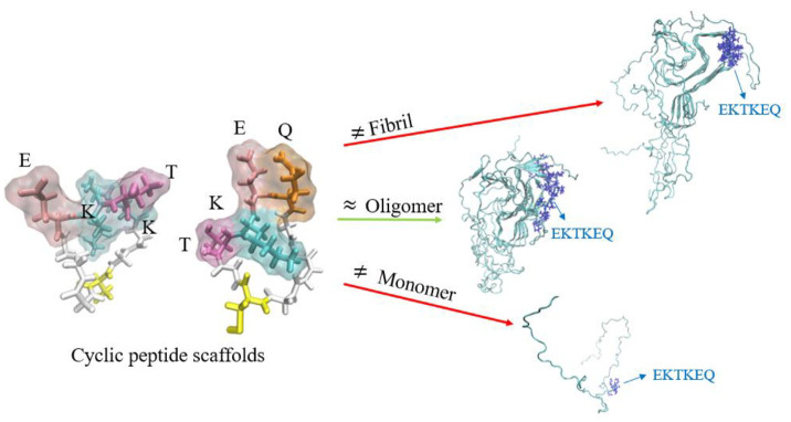
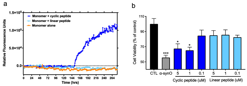
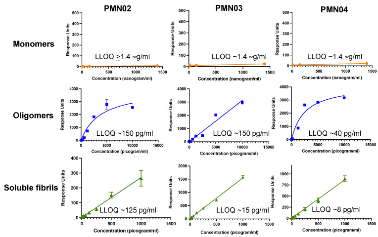
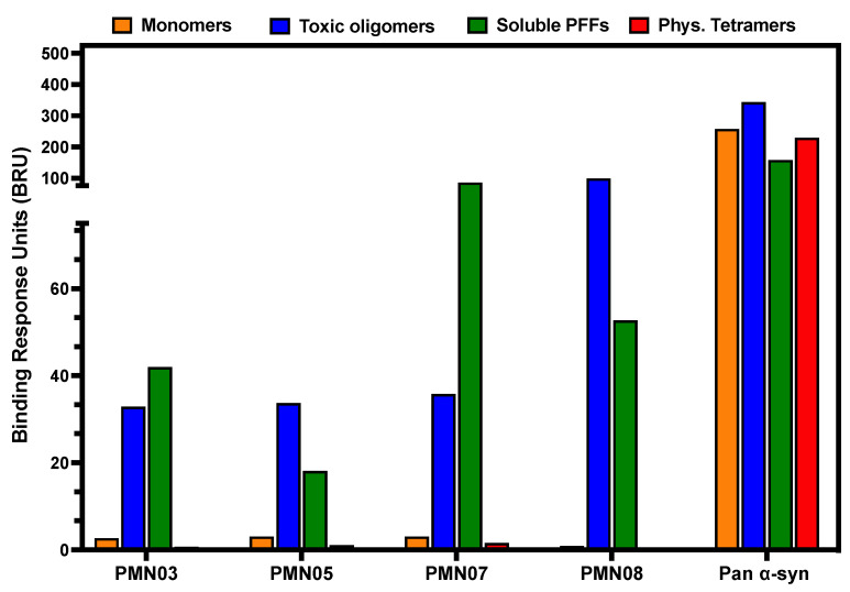
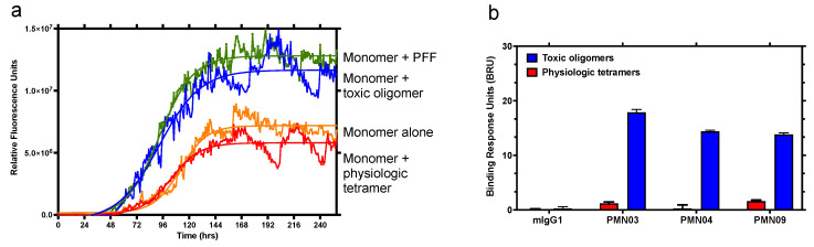
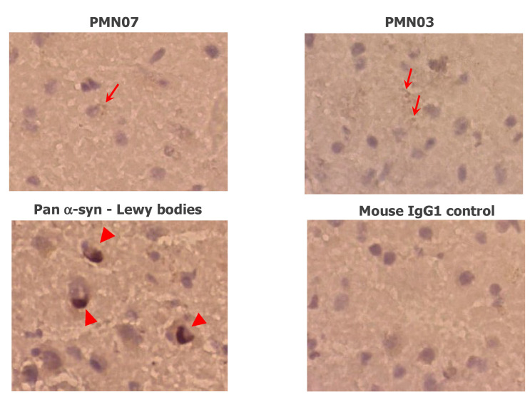
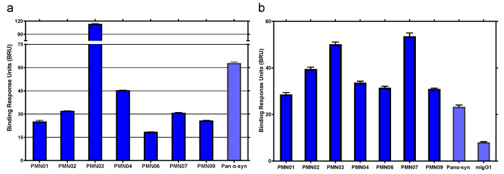
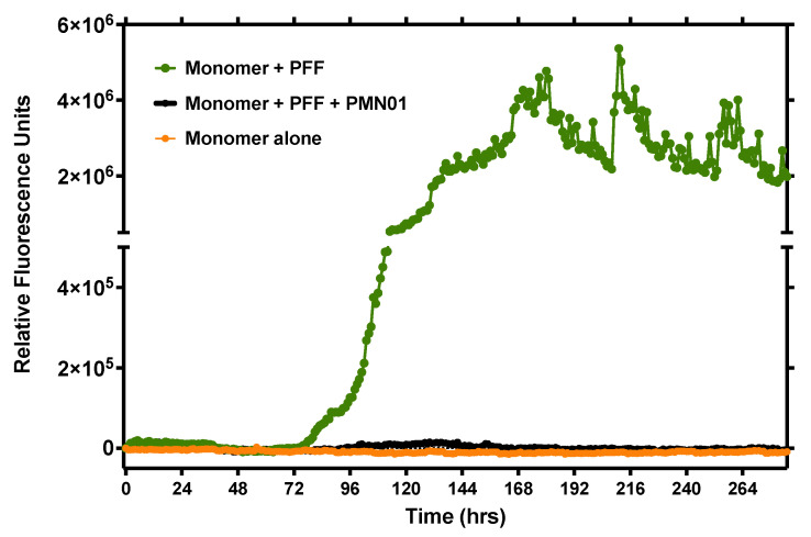
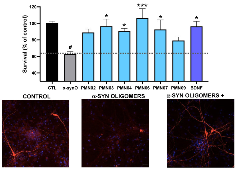
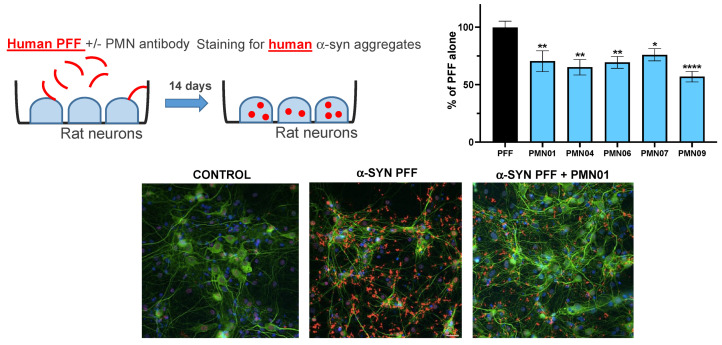
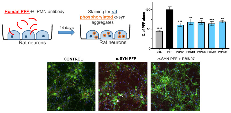
Misfolded toxic forms of alpha-synuclein (α-Syn) have been implicated in the pathogenesis of synucleinopathies, including Parkinson's disease (PD), dementia with Lewy bodies (DLB), and multiple system atrophy (MSA). The α-Syn oligomers and soluble fibrils have been shown to mediate neurotoxicity and cell-to-cell propagation of pathology. To generate antibodies capable of selectively targeting pathogenic forms of α-Syn, computational modeling was used to predict conformational epitopes likely to become exposed on oligomers and small soluble fibrils, but not on monomers or fully formed insoluble fibrils. Cyclic peptide scaffolds reproducing these conformational epitopes exhibited neurotoxicity and seeding activity, indicating their biological relevance. Immunization with the conformational epitopes gave rise to monoclonal antibodies (mAbs) with the desired binding profile showing selectivity for toxic α-Syn oligomers and soluble fibrils, with little or no reactivity with monomers, physiologic tetramers, or Lewy bodies. Recognition of naturally occurring soluble α-Syn aggregates in brain extracts from DLB and MSA patients was confirmed by surface plasmon resonance (SPR). In addition, the mAbs inhibited the seeding activity of sonicated pre-formed fibrils (PFFs) in a thioflavin-T fluorescence-based aggregation assay. In neuronal cultures, the mAbs protected primary rat neurons from toxic α-Syn oligomers, reduced the uptake of PFFs, and inhibited the induction of pathogenic phosphorylated aggregates of endogenous α-Syn. Protective antibodies selective for pathogenic species of α-Syn, as opposed to pan α-Syn reactivity, are expected to provide enhanced safety and therapeutic potency by preserving normal α-Syn function and minimizing the diversion of active antibody from the target by the more abundant non-toxic forms of α-Syn in the circulation and central nervous system.

Keywords: aggregation-based technologies and therapeutics; alpha-synuclein; conformational epitope; fibril; misfolding specific antibody; oligomer; protein aggregation; selectivity; sequence/structure determinants; synucleinopathy.


Conflict of interest statement

N.R.C., J.M.K. and B.Z. are current employees of ProMIS Neurosciences. E.G., S.S.P., S.C.C.H., A.A. and X.P. have received consultation compensation from ProMIS Neurosciences. E.G., S.S.P. and B.Z. possess ProMIS stock options. N.R.C. and J.M.K. possess ProMIS shares and stock options. S.S.P. is the inventor on patent applications for Collective Coordinates computational modeling. S.S.P., N.R.C., X.P. and J.M.K. are inventors on patent applications relating to conformation-specific epitopes in alpha-synuclein, antibodies thereto and methods related thereof. The work presented was financially supported by ProMIS Neurosciences. A.R., J.W., C.S., C.K.Y. and S.-E.N. declare no competing interests.

Figures

Figure 1
Figure 1
Alpha-synuclein residues 57–60 (EKTK) and 59–62 (TKEQ), held in a constrained turn, represent conformational epitopes predicted to be exposed on oligomers and small soluble fibrils but not monomers or fully formed insoluble fibrils. Representative molecular dynamics simulated conformations of the cyclic epitopes with the side chains oriented into solvent are shown.
Figure 2
Figure 2
Conformational cyclic peptide epitopes have seeding activity and toxicity. (a) Aggregation was tracked hourly (shaking for 30 s prior to each reading) with a ThT fluorescent assay after the addition of α-syn monomers alone (100 mM) or in the presence of BSA-conjugated cyclized or linear CGTKEQGGGG peptide (100 nM). Data are representative of 3 independent experiments. (b) Viability of primary rat dopaminergic neurons treated with α-syn oligomers (0.5 μM) or increasing concentrations of BSA-conjugated cyclized or linear peptide. CTL = neurons incubated with vehicle alone. Mean ± SEM of 6 replicates. Global analysis of data performed using one-way ANOVA followed by Dunnett’s multiple comparisons test. * p ≤ 0.05, *** p ≤ 0.001 vs. CTL (vehicle). No statistically significant difference between linear peptide and CTL or between α-syn oligomers and 1, 5 μM cyclic peptide.
Figure 3
Figure 3
Selectivity of mAbs for pathogenic species of α-syn. The binding response of mAbs to various concentrations of α-syn monomers, toxic oligomers and soluble fibrils (sonicated PFFs) was measured in a Millipore immunoassay. Mean ± SD of triplicates shown with the calculated lower limit of quantitation (LLOQ) for each species.
Figure 4
Figure 4
Selective binding of mAbs to pathogenic species of α-syn by SPR. The binding response of immobilized mAbs to α-syn monomers, toxic oligomers, soluble (sonicated) PFFs and physiologic (Phys.) tetramers was measured by SPR. An antibody reactive with all species of α-syn (Pan α-syn) was used as a positive control. The same pattern of binding was observed in 4 independent experiments.
Figure 5
Figure 5
Ability of mAbs to distinguish between toxic oligomers and physiologic tetramers. (a) Aggregation was tracked hourly with a ThT fluorescent assay after the addition of α-syn monomers alone (100 mM) or in the presence of PFF (10 nM), toxic oligomers or physiologic tetramers (both 100 nM). Continuous shaking was used to promote aggregation, giving rise to measurable aggregation with monomers alone. Data are representative of 2 independent experiments. (b) The binding response of immobilized mAbs and negative control mouse IgG1 (mIgG1) to toxic oligomers and physiologic tetramers was measured by SPR. The same pattern of binding was observed in 4 independent experiments. Error bars: SEM.
Figure 6
Figure 6
Reactivity of mAbs with DLB brain sections. mAbs show occasional staining of small aggregates (arrows) as opposed to the dense insoluble deposits of α-syn in Lewy bodies stained by a pan α-syn reactive antibody (arrowheads). No staining is seen with a mouse IgG1 negative control. Images are representative of the staining pattern seen with 17 mAbs tested. Magnification: 40×.
Figure 7
Figure 7
Binding of mAbs to native pathogenic α-syn species in patient brain extract. The binding response of immobilized mAbs to α-syn in brain extractS from DLB (a) and MSA (b) patients was measured by SPR. A pan α-syn reactive antibody and mouse IgG1 (mIgG1) were used as controls. Results shown are the mean ± SEM of 2 (a) or 4 (b) independent studies.
Figure 8
Figure 8
mAb inhibition of PFF seeding activity. Aggregation was tracked hourly with a ThT fluorescent assay (shaking for 30 sec prior to each reading) after the addition of α-syn monomers alone (100 mM) or in the presence of sonicated human PFF (10 nM) as a seeding agent, without or with mAb (0.1 nM). Data are representative of 2 independent experiments.
Figure 9
Figure 9
mAb inhibition of oligomer toxicity in dopaminergic neurons. Cultures of primary rat dopaminergic neurons were exposed to toxic α-syn oligomers (α-synO; 0.5 μM) without or with mAbs (0.25 μM). Survival is expressed as the percentage of viable neurons compared to a control culture with vehicle only (CTL). BDNF was used as a positive control. Results shown are the mean ± SEM of 6 replicate cultures. A global analysis of the data was performed using one-way ANOVA followed by Dunnett’s multiple comparisons test. # p < 0.005 vs. CTL, * p ≤ 0.05 vs. α-synO, *** p ≤ 0.001 vs. α-synO. No statistically significant difference between BDNF and CTL or mAbs and CTL. Representative images of cultures with staining for neurons (MAP2, red) and nuclei (blue) are shown.
Figure 10
Figure 10
mAb inhibition of PFF uptake and intracellular aggregate formation. Cultures of primary rat hippocampal neurons were exposed to sonicated human PFF (1 μg/mL) without or with mAbs (0.05 μM, except for PMN09 at 0.25 μM). Cultures were stained 14 days later for neuronal marker MAP2 (green), aggregates of human α-syn (red) and cell nuclei (blue). Results are expressed as a percentage of the human α-syn staining area with PFF alone and show the mean ± SEM of 6 replicate cultures. A global analysis of the data was performed using one-way ANOVA followed by Dunnett’s multiple comparisons test. * p ≤ 0.05, ** p ≤ 0.01, **** p ≤ 0.0001 vs. PFF.
Figure 11
Figure 11
mAb inhibition of the recruitment of endogenous rat α-syn into phosphorylated aggregates. Cultures of primary rat hippocampal neurons were exposed to sonicated human PFF (1 μg/mL) without or with mAbs (0.05 μM, except for PMN09 at 0.25 μM). CTL = neurons incubated with vehicle alone. Cultures were stained 14 days later for neuronal marker MAP2 (green), aggregates of phosphorylated rat α-syn (red) and cell nuclei (blue). Results are expressed as a percentage of the phosphorylated rat α-syn staining area with PFF alone and show the mean ± SEM of 6 replicate cultures. A global analysis of the data was performed using one-way ANOVA followed by Dunnett’s multiple comparisons test. ** p ≤ 0.01, *** p ≤ 0.001, **** p ≤ 0.0001 vs. PFF.

References

    1. Brundin P., Dave K.D., Kordower J.H. Therapeutic approaches to target alpha-synuclein pathology. Exp. Neurol. 2017;298:225–235. doi: 10.1016/j.expneurol.2017.10.003. - DOI - PMC - PubMed
    1. Lashuel H.A., Overk C.R., Oueslati A., Masliah E. The many faces of alpha-synuclein: From structure and toxicity to therapeutic target. Nat. Rev. Neurosci. 2013;14:38–48. doi: 10.1038/nrn3406. - DOI - PMC - PubMed
    1. Fusco G., Chen S.W., Williamson P.T.F., Cascella R., Perni M., Jarvis J.A., Cecchi C., Vendruscolo M., Chiti F., Cremades N., et al. Structural basis of membrane disruption and cellular toxicity by alpha-synuclein oligomers. Science. 2017;358:1440–1443. doi: 10.1126/science.aan6160. - DOI - PubMed
    1. Bengoa-Vergniory N., Roberts R.F., Wade-Martins R., Alegre-Abarrategui J. Alpha-synuclein oligomers: A new hope. Acta Neuropathol. 2017;134:819–838. doi: 10.1007/s00401-017-1755-1. - DOI - PMC - PubMed
    1. Plotkin S.S., Cashman N.R. Passive immunotherapies targeting Abeta and tau in Alzheimer’s disease. Neurobiol. Dis. 2020;144:105010. doi: 10.1016/j.nbd.2020.105010. - DOI - PMC - PubMed

LinkOut - more resources