. 2022 Sep 14;10(9):2275.
doi: 10.3390/biomedicines10092275.

The Adipose Organ Is a Unitary Structure in Mice and Humans

Affiliations

The Adipose Organ Is a Unitary Structure in Mice and Humans

A Giordano et al. Biomedicines. .

Abstract

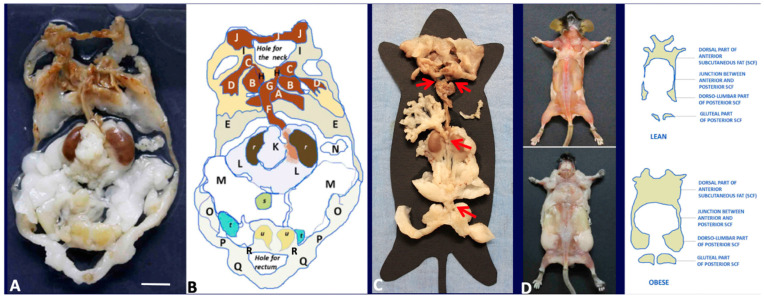
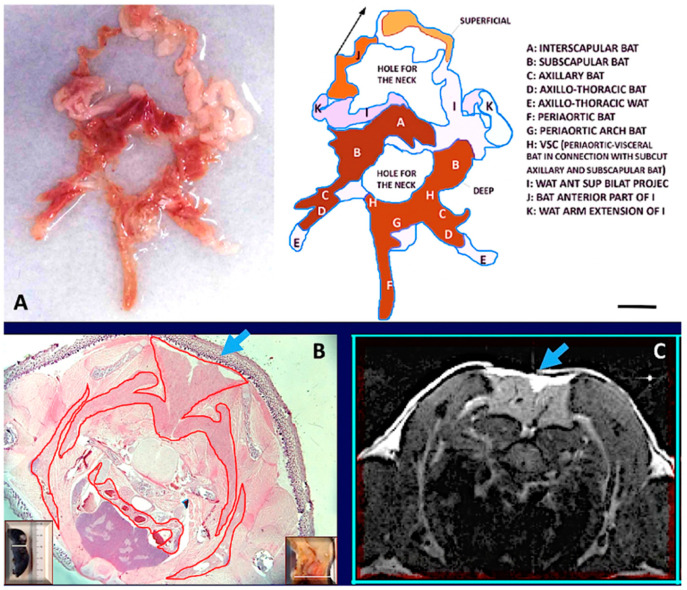
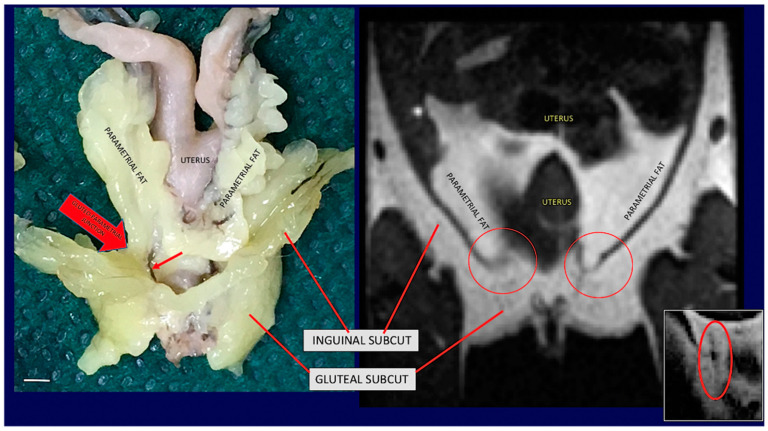
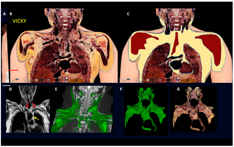
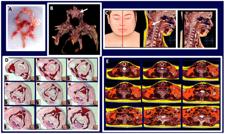
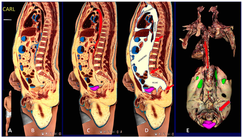
Obesity is the fifth leading cause of death worldwide. In mice and humans with obesity, the adipose organ undergoes remarkable morpho-functional alterations. The comprehension of the adipose organ function and organization is of paramount importance to understand its pathology and formulate future therapeutic strategies. In the present study, we performed anatomical dissections, magnetic resonance imaging, computed axial tomography and histological and immunohistochemical assessments of humans and mouse adipose tissues. We demonstrate that most of the two types of adipose tissues (white, WAT and brown, BAT) form a large unitary structure fulfilling all the requirements necessary to be considered as a true organ in both species. A detailed analysis of the gross anatomy of mouse adipose organs in different pathophysiological conditions (normal, cold, pregnancy, obesity) shows that the organ consists of a unitary structure composed of different tissues: WAT, BAT, and glands (pregnancy). Data from autoptic dissection of 8 cadavers, 2 females and 6 males (Age: 37.5 ± 9.7, BMI: 23 ± 2.7 kg/m2) and from detailed digital dissection of 4 digitalized cadavers, 2 females and 2 males (Age: 39 ± 14.2 years, BMI: 22.8 ± 4.3 kg/m2) confirmed the mixed (WAT and BAT) composition and the unitary structure of the adipose organ also in humans. Considering the remarkable endocrine roles of WAT and BAT, the definition of the endocrine adipose organ would be even more appropriate in mice and humans.

Keywords: beige adipocyte; brown adipocyte; mammary gland; obesity; organ; subcutaneous fat; visceral fat; white adipocyte.


Conflict of interest statement

The authors declare no conflict of interest.

Figures

Figure 1
Figure 1
Gross anatomy of mouse adipose organ in different experimental conditions. (A): Anatomical preparation of the adipose organ as a unitary structure (adult, male C57BL/6 mouse, normoweight). Subcutaneous-visceral continuities (SVCs) are well visible and indicated in the diagram legend. Preputial glands and kidneys are left to facilitate anatomical orientation. White (WAT) and brown (BAT) adipose tissues are recognizable by their specific colors. (B): Legend: A: interscapular BAT (subcutaneous, SC), B: subscapular BAT (SC), C: supraclavicular BAT (SC), D: axillary BAT (SC), E: axillo-thoracic WAT (SC), F and G: periaortic mediastinal BAT (Visceral, V), H: SVC, I: cervical subcutaneous WAT (SC), J: cervical subcutaneous BAT (SC), K: mesenteric WAT (V), L: perirenal-retroperitoneal WAT-BAT (V), M: epididymal WAT (V), N: omental WAT (V), O: dorso-lumbar WAT (SC), P: inguinal WAT (SC), Q: gluteal WAT (SC). A + B + C + D + E + I + J: anterior subcutaneous region of adipose organ. O + P + Q: posterior subcutaneous region of the adipose organ. F + G + K + L + M + N: visceral region of the adipose organ. H and R: SVCs. r: kidney, s: urinary bladder, u: preputial glands. (C): Anatomical preparation of the adipose organ (adult, female C57BL/6 mouse, normoweight) as a unitary structure laying on a template. Red arrows point to SVCs (H and R in (B)) and mesenteric-abdominopelvic (inter-visceral) connection. (D): Dorsal view of adult C57BL6 female mice after skin removal. Anterior and posterior subcutaneous parts of the adipose organ are visible in both normoweight (upper panel) and obese animals. Bar: 8 mm in (A,B), 13 mm in (C) and 40 mm in (D).
Figure 2
Figure 2
The fat neckerchief of mouse Adipose Organ. (A): Anatomical preparation of fat neckerchief displaying its continuity with peri-aortic mediastinal fat (adult C57BL/6 at room temperature). The superficial white fat of the neckerchief is separated and folded up as indicated by the arrow in the diagram. (B): Histology (newborn, C57BL/6) and axial T1-weighted MRI image (adult, C57BL/6) (in (C)) of the interscapular region, show the continuity between interscapular fat depot with the axillary fat, outlined by the red line surrounding brown fat in histology. Further evidence is shown by serial sections in histology and MRI (Supplementary Figure S7). The interscapular region (indicated by light blue arrows) also shows that brown (identified by a hypointense area) and white adipose tissue (hyperintense area) are contained in the same depot as also evident in anatomical preparations and histology. Insets in (B) show the plane of the section in the whole animal (left) and in its sagittal section (right). Bar: 10 mm in (A,B), and 1.5 mm in (C).
Figure 3
Figure 3
Gross anatomy and MRI of subcuteous-visceral continuity in the lower part of mouse Adipose Organ. (Left): Anatomical preparation of an adult C57BL/6 mouse showing subcutaneous-visceral continuity between parametrial and gluteal fat. (Right): coronal T1-weighted MRI of the same areas shown in the left panel of the same animal in vivo. Anatomical continuity is indicated in the visceral (parametrial) and subcutaneous (inguinal) fat by arrow and red circles. A small red arrow on the left panel shows a vessel in the junction. A similar structure was also detected by MRI (red circled area in small, squared panel on the right). Bar 4.5 mm in both panels.
Figure 4
Figure 4
Gross anatomy and MRI of subcutaneous-visceral continuity in human Adipose Organ. (A): Oblique frontal section of a digital cadaver (Vicky). (B): Supraclavicular-mediastinal continuity of the section shown in (A). (C): The same section shown in (B), with the fat area highlighted in yellow. (D): Routine MRI in an adult male. Red arrows indicate subcutaneous-visceral fat connection (SVC) and the yellow arrow indicates the mediastinal fat. (E): 3D rendering (green) of the SVC in a 10-year-old boy. (F,G): 3D reconstruction of the fat present in the supraclavicular-mediastinal continuity shown in (B,C). Fat area is highlighted in three planes (axial, coronal, and sagittal); in F fat is in green for comparison with (E); in (G) fat is shown in its original colors. Bar: 60 mm in (B,C), 45 mm in (F,G).
Figure 5
Figure 5
Comparative data between mouse and human fat neckerchief. (A): Human fat neckerchief. (B): 3D reconstruction of the fat areas obtained through the entire set of Vicky’s cervical sections. Dorsal view, the deep neck brown fat mass is indicated by the arrow. (C): Vicky median sagittal section (left panel) in which the subcutaneous fat is highlighted in yellow and the brown fat in orange (central panel). The arrow indicates the deep neck brown fat also shown in (B). The right panel shows the original section with no highlights. (D,E) show a comparison between the brown fat forming the neckerchief in a mouse and in Vicky serial sections (original sections are shown in Supplementary Figures S7 and S15). Bars: 15 mm in (A), 60 mm in (B), 14 cm in (C), 15 mm in (D), 6 cm in (E).
Figure 6
Figure 6
Digital cadaver section and 3D reconstruction showing upper and lower subcutaneous visceral continuity in the Adipose Organ. (A): Sagittal oblique section of the digital cadaver, Carl. (BD): Mediastinal-abdomino-pelvic and gluteal subcutaneous-visceral connection (SVC). (B): original section, (C): aorta and upper mesenteric artery in red, (D): fat highlighted in white. Arrows in (D) point to mediastinic-abdominal (upper), abdomino-pelvic (middle) and gluteal-pelvic (lower) connections. Gluteal-pelvic SVC is also shown in Supplementary Figure S16. Panel (E) shows a 3D reconstruction of the whole visceral fat. Dorsal view in which the continuity between retroperitoneal and pelvic fat surrounding the urinary bladder is more evident, indicated by the arrow. Green: kidneys (parts of dorsal surface not covered by retroperitoneal fat), violet: urinary bladder. Arrows point to periaortic fat in the mediastinic-abdominal connection. See also Suppl movie. Bar: in (BD) 60 mm, in (E) 120 mm.
Figure 7
Figure 7
2D and 3D reconstructions of human Adipose Organ. Digital cadaver Vicky. (A): Representative images showing the whole adipose organ in a coronal section, (B): the 3D reconstruction of isolated visceral fat in connection with the supraclavicular-axillary part of the adipose organ, and (C): the visceral (brown, beige, and pale white) and subcutaneous (yellow) parts of the adipose organ (still image from S Movie 1). Bar: 65 mm in (A), 35 mm in (B) and 75 mm in (C).

References

    1. Cinti S. Adipose Organ Development and Remodeling. Compr. Physiol. 2018;8:1357–1431. doi: 10.1002/cphy.c170042. - DOI - PubMed
    1. Cannon B., Nedergaard J. Brown adipose tissue: Function and physiological significance. Physiol Rev. 2004;84:277–359. doi: 10.1152/physrev.00015.2003. - DOI - PubMed
    1. Barbatelli G., Murano I., Madsen L., Hao Q., Jimenez M., Kristiansen K., Giacobino J.P., De Matteis R., Cinti S. The emergence of cold-induced brown adipocytes in mouse white fat depots is determined predominantly by white to brown adipocyte transdifferentiation. Am. J. Physiol. Endocrinol. Metab. 2010;298:E1244–E1253. doi: 10.1152/ajpendo.00600.2009. - DOI - PubMed
    1. Altshuler-Keylin S., Shinoda K., Hasegawa Y., Ikeda K., Hong H., Kang Q., Yang Y., Perera R.M., Debnath J., Kajimura S. Beige Adipocyte Maintenance Is Regulated by Autophagy-Induced Mitochondrial Clearance. Cell Metab. 2016;24:402–419. doi: 10.1016/j.cmet.2016.08.002. - DOI - PMC - PubMed
    1. Kotzbeck P., Giordano A., Mondini E., Murano I., Severi I., Venema W., Cecchini M.P., Kershaw E.E., Barbatelli G., Haemmerle G., et al. Brown adipose tissue whitening leads to brown adipocyte death and adipose tissue inflammation. J. Lipid Res. 2018;59:784–794. doi: 10.1194/jlr.M079665. - DOI - PMC - PubMed

LinkOut - more resources