Review
. 2022 Sep 16;12(9):2235.
doi: 10.3390/diagnostics12092235.

Artificial Intelligence in Orthopedic Radiography Analysis: A Narrative Review

Affiliations
Review

Artificial Intelligence in Orthopedic Radiography Analysis: A Narrative Review

Kenneth Chen et al. Diagnostics (Basel). .

Abstract

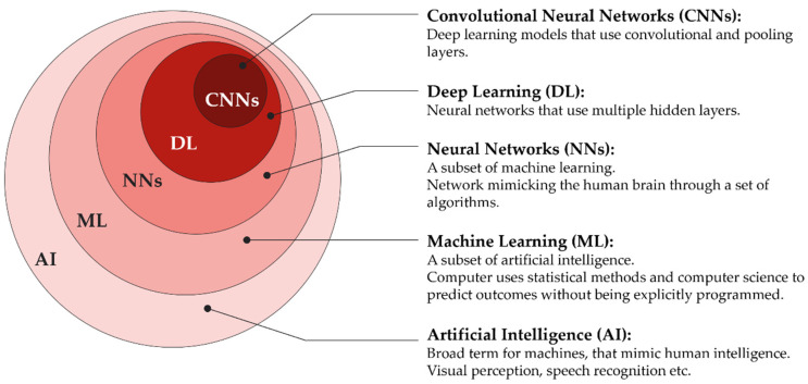
Artificial intelligence (AI) in medicine is a rapidly growing field. In orthopedics, the clinical implementations of AI have not yet reached their full potential. Deep learning algorithms have shown promising results in computed radiographs for fracture detection, classification of OA, bone age, as well as automated measurements of the lower extremities. Studies investigating the performance of AI compared to trained human readers often show equal or better results, although human validation is indispensable at the current standards. The objective of this narrative review is to give an overview of AI in medicine and summarize the current applications of AI in orthopedic radiography imaging. Due to the different AI software and study design, it is difficult to find a clear structure in this field. To produce more homogeneous studies, open-source access to AI software codes and a consensus on study design should be aimed for.

Keywords: X-ray; artificial intelligence; deep learning; machine learning; musculoskeletal imaging; radiograph.


Conflict of interest statement

The authors declare no conflict of interest.

Figures

Figure 1
Figure 1
Number of PubMed search results for “artificial intelligence” with an exponential growth in numbers of published articles after 2016.
Figure 2
Figure 2
A schematic illustration of the role of artificial intelligence (AI) in orthopedic radiography analysis. AI can play a role in supporting and enhancing image interpretation, research and development, as well as decision-making for treatment plans.
Figure 3
Figure 3
Set diagram describing the relationship of Artificial intelligence (AI) being the umbrella term for everything inside the outer circle, Machine learning (ML) for everything inside the second-outer circle, etc. Neural networks (NN), deep learning (DL), and convolutional neural networks (CNNs).
Figure 4
Figure 4
Illustration describing the mechanism of clusterization: In the left image, no specific order can be seen. For clusterization, different shapes are recognized and organized in a group (cluster) as seen in the right image. Shapes that do not belong in any group (e.g., stars) are sorted out. This can help ML models to recognize patterns that would otherwise be difficult to recognize.
Figure 5
Figure 5
Max pooling layer: the input layer (left) is filtered by a 2 × 2 max pooling layer computing the output with the highest activation (number) of the filtered area in the preceding layer into a smaller output layer. One can imagine a 2 × 2 filter starting on the top left: The first filtered area will be the blue area. The number six, being the highest, will be computed as 1 × 1 in the output, representing the 2 × 2 input. After that, the filter will go to the next area that has not been filtered, e.g., two cells to the right, and repeat the process. With filtering methods like this, the required computational power can be reduced.
Figure 6
Figure 6
Internal vs. external validation. Internal validation uses one dataset and splits it into training and validation, the ML model is then trained and validated on the same dataset resulting in higher risk of overfitting. External validation uses one dataset for training and a meaningfully different dataset for validation. When (geographically) different datasets are used, the risk of overfitting can be significantly reduced.

References

    1. Shen D., Wu G., Suk H.-I. Deep Learning in Medical Image Analysis. Annu. Rev. Biomed. Eng. 2017;19:221–248. doi: 10.1146/annurev-bioeng-071516-044442. - DOI - PMC - PubMed
    1. McCarthy J., Minsky M.L., Rochester N., Shannon C.E. A Proposal for the Dartmouth Summer Research Project on Artificial Intelligence, August 31, 1955. AI Mag. 2006;27:12. doi: 10.1609/aimag.v27i4.1904. - DOI
    1. Hamet P., Tremblay J. Artificial intelligence in medicine. Metabolism. 2017;69S:S36–S40. doi: 10.1016/j.metabol.2017.01.011. - DOI - PubMed
    1. Goodfellow I., Bengio Y., Courville A. Deep Learning. The MIT Press; Cambridge, MA, USA: 2016.
    1. Rouzrokh P., Wyles C.C., Philbrick K.A., Ramazanian T., Weston A.D., Cai J.C., Taunton M.J., Lewallen D.G., Berry D.J., Erickson B.J., et al. A Deep Learning Tool for Automated Radiographic Measurement of Acetabular Component Inclination and Version After Total Hip Arthroplasty. J. Arthroplast. 2021;36:2510–2517.e6. doi: 10.1016/j.arth.2021.02.026. - DOI - PMC - PubMed

LinkOut - more resources