

Acute Effects of Static and Proprioceptive Neuromuscular Facilitation Stretching of the Plantar Flexors on Ankle Range of Motion and Muscle-Tendon Behavior in Children with Spastic Cerebral Palsy-A Randomized Clinical Trial
- PMID: 36141875
- PMCID: PMC9517397
- DOI: 10.3390/ijerph191811599
Acute Effects of Static and Proprioceptive Neuromuscular Facilitation Stretching of the Plantar Flexors on Ankle Range of Motion and Muscle-Tendon Behavior in Children with Spastic Cerebral Palsy-A Randomized Clinical Trial
Abstract
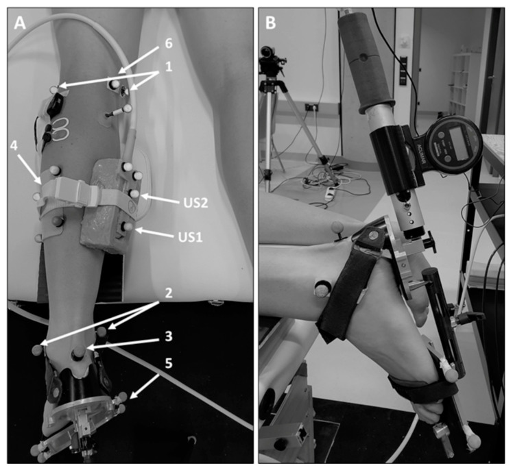
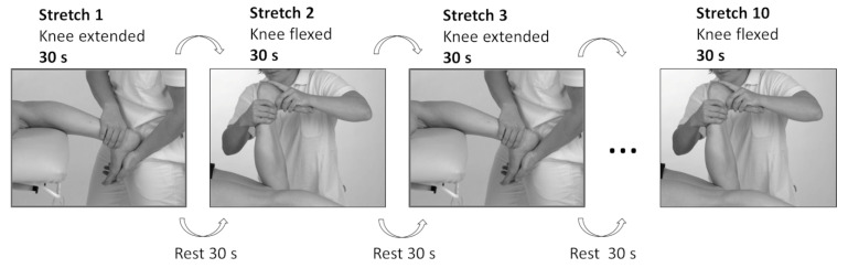
Stretching is considered a clinically effective way to prevent muscle contracture development in children with spastic cerebral palsy (CP). Therefore, in this study, we assessed the effects of a single session of proprioceptive neuromuscular facilitation (PNF) or static stretching (SS) on ankle joint range of motion (RoM) and gastrocnemius muscle-tendon behavior in children with CP. During the SS (n = 8), the ankle joint was held in maximum dorsiflexion (30 s). During the PNF stretching (n = 10), an isometric contraction (3-5 s) was performed, followed by stretching (~25 s). Ten stretches were applied in total. We collected data via dynamometry, 3D motion capture, 2D ultrasound, and electromyography, before and after the stretching sessions. A mixed ANOVA was used for the statistical analysis. Both ankle RoM and maximum dorsiflexion increased over time (F(1,16) = 7.261, p < 0.05, η² = 0.312; and F(1,16) = 4.900, p < 0.05, η² = 0.234, respectively), without any difference between groups. An interaction effect (F(1,12) = 4.768, p = 0.05, η² = 0.284) was observed for muscle-tendon unit elongation (PNF: -8.8%; SS: +14.6%). These findings suggest a positive acute effect of stretching on ankle function. However, SS acutely increased muscle-tendon unit elongation, while this decreased after PNF stretching, indicating different effects on the spastic muscles. Whether PNF stretching has the potential to cause positive alterations in individuals with CP should be elucidated in future studies.
Keywords: Achilles tendon; hold-relax PNF stretching; muscle activity; paresis; plantar flexors; spasticity.
Conflict of interest statement
The authors declare no conflict of interest. The funders had no role in the design of the study, in the collection, analyses, or interpretation of data in the writing of the manuscript, or in the decision to publish the results.
Figures
References
Publication types
MeSH terms
LinkOut - more resources
Full Text Sources
Medical
Miscellaneous
