. 2022 Sep 6;23(18):10219.
doi: 10.3390/ijms231810219.

Icariin Alleviates Escherichia coli Lipopolysaccharide-Mediated Endometritis in Mice by Inhibiting Inflammation and Oxidative Stress

Affiliations

Icariin Alleviates Escherichia coli Lipopolysaccharide-Mediated Endometritis in Mice by Inhibiting Inflammation and Oxidative Stress

Aftab Shaukat et al. Int J Mol Sci. .

Abstract

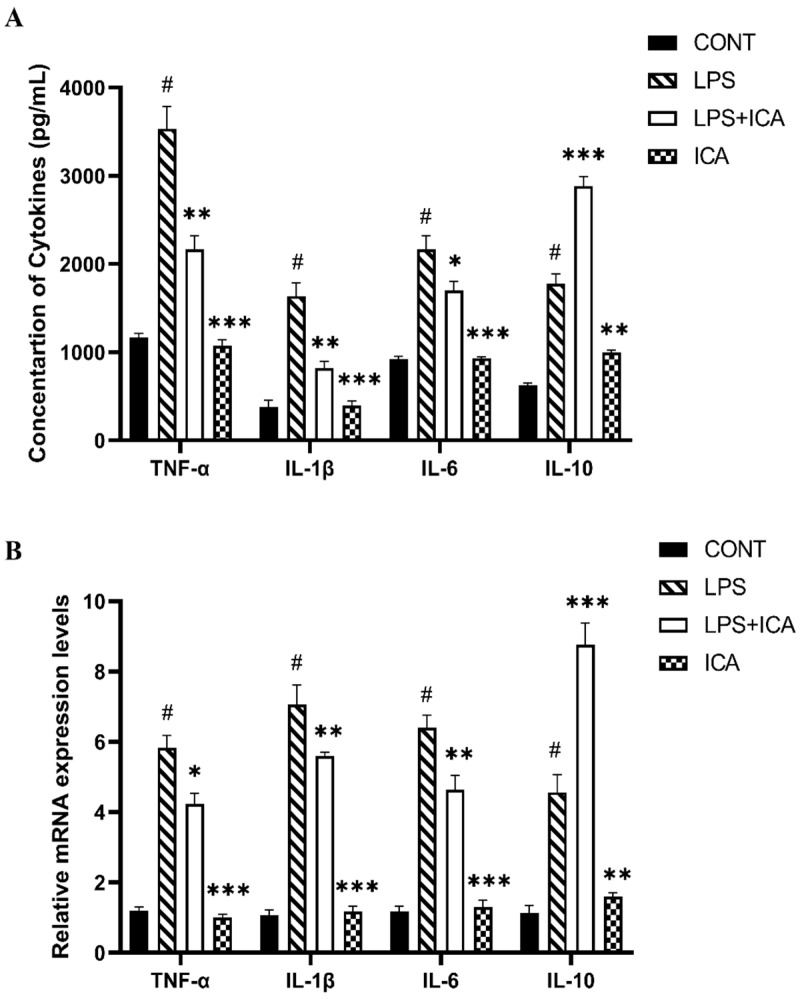
Icariin (ICA) is a naturally occurring phytochemical agent primarily extracted from Epimedium Brevicornum Maxim (Family Berberidaceae) with a broad spectrum of bioactivities. Endometritis is a uterine disease that causes enormous losses in the dairy industry worldwide. In this study, anti-inflammatory and anti-oxidant properties of ICA were investigated against lipopolysaccharide (LPS)-induced endometritis in mice to investigate possible underlying molecular mechanisms. Sixty heathy female Kunming mice were randomly assigned to four groups (n = 15), namely control, LPS, LPS + ICA, and ICA groups. The endometritis was induced by intrauterine infusion of 50 µL of LPS (1 mg/mL). After 24 h of onset of LPS-induced endometritis, ICA groups were injected thrice by ICA intraperitoneally six hours apart. Histopathological examination, enzyme linked immunosorbent assay (ELISA), real time quantitative polymerase chain reaction (RT-qPCR), western blotting, and immunohistochemistry were used in this study. Histological alterations revealed that ICA markedly mitigated uterine tissue injury caused by LPS. The results showed that the ICA inhibited the production of pro-inflammatory cytokines (IL-1ß, IL-6, and TNF-α) and boosted the production of anti-inflammatory cytokines (IL-10). Additionally, ICA modulated the expression of malondialdehyde (MDA), reactive oxygen species (ROS), superoxide dismutase 1 (SOD1), catalase (CAT), and glutathione peroxidase 1 (Gpx1) induced by LPS. The administration of ICA significantly (p < 0.05) improved the mRNA and protein expression of Toll-like receptor (TLR) 4. The western blotting and ELISA finding revealed that the ICA repressed LPS-triggered NF-κB pathway activation. Moreover, ICA improved the antioxidant defense system via activation of the Nrf2 pathway. The results revealed that ICA up-regulated the mRNA and protein expression of Nuclear erythroid-2-related factor (Nrf2), NAD(P)H: quinone oxidoreductase 1 (NQO1), heme oxygenase-1 (HO-1), and glutamate-cysteine ligase catalytic subunit (GCLC) under LPS exposure. Conclusively, our findings strongly suggested that ICA protects endometritis caused by LPS by suppressing TLR4-associated NF-κB and Nrf2 pathways. Altogether, these innovative findings may pave the way for future studies into the therapeutic application of ICA to protect humans and animals against endometritis.

Keywords: NF-κB and Nrf2 pathway; TLR4; endometritis; icariin; lipopolysaccharide.


Conflict of interest statement

The authors declared no conflicts of interest.

Figures

Figure 1
Figure 1
(A) Epimedium Brevicornum. (B) Structure of ICA. (C) HPLC chromatogram of ICA (the purity of ICA was more than or equal to 98%).
Figure 2
Figure 2
The effect of ICA on LPS-induced uterine injury in mice (A) H&E analysis of uterine tissue, Scale bar: 100 μm. (B) Histopathological scoring of the murine uterus. (C) W/D ratio. (D) MPO activity assay and (E) NO concentration. The data were statistically presented as means ± SEM. The # p < 0.001 between CONT and LPS groups, and * p < 0.05, ** p < 0.01, *** p < 0.001 between LPS and ICA therapy groups. (CONT, LPS, and ICA stand for control, lipopolysaccharide, and Icariin groups [The ICA dose used was 50 mg/kg for uterine tissue]).
Figure 3
Figure 3
Effect of ICA in LPS-triggered secretion of cytokines. (A) Concentration of TNF-α (pg/mL), IL-1β (pg/mL), IL-6 (pg/mL) and IL-10 (pg/mL). (B) The relative mRNA expression levels of TNF-α, IL-1β, IL-6, and IL-10. The data were statistically presented as means ± SEM. The # p < 0.001 between CONT and LPS groups, and * p < 0.05, ** p < 0.01, *** p < 0.001 between LPS and ICA therapy groups. (CONT, LPS, and ICA stand for control, lipopolysaccharide, and Icariin groups. [The ICA dose used was 50 mg/kg for uterine tissue]).
Figure 4
Figure 4
Consequences of ICA on LPS-induced TLR4 expression. (A) The immunohistochemical expression of TLR4 protein. (B) The relative mRNA expression level of the TLR4 gene. The data were statistically presented as means ± SEM. The # p < 0.001 between CONT and LPS groups, and ** p < 0.01, *** p < 0.001 between LPS and ICA therapy groups. (CONT, LPS and ICA stand for control, lipopolysaccharide and Icariin groups [The ICA dose used was 50 mg/kg for uterine tissue]).
Figure 5
Figure 5
Outcomes of ICA in LPS-induced expression NF-κB signaling pathway. (A) The expression levels of the NF-κB-p65 and its phosphorylated (p-NF-κB-p65) form were detected by ELISA. (B) The expression levels of total IκBα and its phosphorylated (p-IκBα) form were detected by ELISA. (C) The protein expression levels of the TLR4, phosphorylated (p-NF-κB-p65), and phosphorylated (p-IκBα) were detected by western blotting. (D) The quantification of all the analyzed proteins. The β-Actin was used as a control. The data were statistically presented as means ± SEM. The # p < 0.001 between CONT and LPS groups, and * p < 0.05, ** p < 0.01, *** p < 0.001 between LPS and ICA therapy groups. (CONT, LPS, and ICA stand for control, lipopolysaccharide, and Icariin groups [The ICA dose used was 50 mg/kg for uterine tissue]).
Figure 6
Figure 6
Influence of ICA treatment on LPS-induced oxidative stress-related genes via activation of Nrf2 pathway in murine uterus. (A) The relative mRNA expression levels of SOD1, CAT and Gpx1 was determined by RT-qPCR. (B) The relative mRNA expression levels of Nrf2, HO-1, NQO1, and GCLC. (C) The protein expressions of Nrf2, HO-1, and NQO1 were detected by western blotting. (D) The quantification of all the analyzed proteins. The data were statistically presented as means ± SEM. The # p < 0.001 between CONT and LPS groups, and * p < 0.05, ** p < 0.01, *** p < 0.001 between LPS and ICA therapy groups. (CONT, LPS, Nrf2, HO-1, NQO1, GCLC, and ICA stand for control, lipopolysaccharide, Nuclear erythroid-2-related factor, heme oxygenase-1, NAD(P)H: quinone oxidoreductase 1, glutamate-cysteine ligase catalytic subunit, and Icariin. [The ICA dose used was 50 mg/kg for uterine tissue]).

References

    1. Jiang K., Guo S., Yang J., Liu J., Shaukat A., Zhao G., Wu H., Deng G. Matrine alleviates Staphylococcus aureus lipoteichoic acid-induced endometritis via suppression of TLR2-mediated NF-κB activation. Int. Immunopharmacol. 2019;70:201–207. doi: 10.1016/j.intimp.2019.02.033. - DOI - PubMed
    1. Wu H., Zhao G., Jiang K., Li C., Qiu C., Deng G. Engeletin alleviates lipopolysaccharide-induced endometritis in mice by inhibiting TLR4-mediated NF-κB activation. J. Agric. Food Chem. 2016;64:6171–6178. doi: 10.1021/acs.jafc.6b02304. - DOI - PubMed
    1. Liu J., Guo S., Jiang K., Zhang T., Zhiming W., Yaping Y., Jing Y., Shaukat A., Deng G. miR-488 mediates negative regulation of the AKT/NF-κB pathway by targeting Rac1 in LPS-induced inflammation. J. Cell. Physiol. 2020;235:4766–4777. doi: 10.1002/jcp.29354. - DOI - PubMed
    1. Wu H., Jiang K., Yin N., Ma X., Zhao G., Qiu C., Deng G. Thymol mitigates lipopolysaccharide-induced endometritis by regulating the TLR4- and ROS-mediated NF-kappaB signaling pathways. Oncotarget. 2017;8:20042–20055. doi: 10.18632/oncotarget.15373. - DOI - PMC - PubMed
    1. Lv X., Fu K., Li W., Wang Y., Wang J., Li H., Tian W., Cao R. TIIA attenuates LPS-induced mouse endometritis by suppressing the NF-κB signaling pathway. Can. J. Physiol. Pharmacol. 2015;93:967–971. doi: 10.1139/cjpp-2015-0003. - DOI - PubMed

MeSH terms

LinkOut - more resources