. 2022 Sep 6;23(18):10235.
doi: 10.3390/ijms231810235.

TNF-α and IL-1β Modulate Blood-Brain Barrier Permeability and Decrease Amyloid-β Peptide Efflux in a Human Blood-Brain Barrier Model

Affiliations

TNF-α and IL-1β Modulate Blood-Brain Barrier Permeability and Decrease Amyloid-β Peptide Efflux in a Human Blood-Brain Barrier Model

Romain Versele et al. Int J Mol Sci. .

Abstract

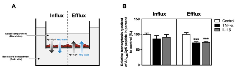
The blood-brain barrier (BBB) is a selective barrier and a functional gatekeeper for the central nervous system (CNS), essential for maintaining brain homeostasis. The BBB is composed of specialized brain endothelial cells (BECs) lining the brain capillaries. The tight junctions formed by BECs regulate paracellular transport, whereas transcellular transport is regulated by specialized transporters, pumps and receptors. Cytokine-induced neuroinflammation, such as the tumor necrosis factor-α (TNF-α) and interleukin-1β (IL-1β), appear to play a role in BBB dysfunction and contribute to the progression of Alzheimer's disease (AD) by contributing to amyloid-β (Aβ) peptide accumulation. Here, we investigated whether TNF-α and IL-1β modulate the permeability of the BBB and alter Aβ peptide transport across BECs. We used a human BBB in vitro model based on the use of brain-like endothelial cells (BLECs) obtained from endothelial cells derived from CD34+ stem cells cocultivated with brain pericytes. We demonstrated that TNF-α and IL-1β differentially induced changes in BLECs' permeability by inducing alterations in the organization of junctional complexes as well as in transcelluar trafficking. Further, TNF-α and IL-1β act directly on BLECs by decreasing LRP1 and BCRP protein expression as well as the specific efflux of Aβ peptide. These results provide mechanisms by which CNS inflammation might modulate BBB permeability and promote Aβ peptide accumulation. A future therapeutic intervention targeting vascular inflammation at the BBB may have the therapeutic potential to slow down the progression of AD.

Keywords: Alzheimer’s disease; IL-1β; TNF-α; amyloid-β peptide; blood-brain barrier; inflammation.


Conflict of interest statement

The authors declare no conflict of interest.

Figures

Figure 1
Figure 1
BLECs’ response to TNF-α and IL-1β treatment. (A) After 24 h, the effects of 10 ng·mL−1 of TNF-α or IL-1β on cell viability were performed using MTT assay. A 10% DMSO treatment was used as positive control of cell death. (B) The proinflammatory markers (VCAM-1, NLRP3 and ICAM-1) after TNF-α or IL-1β treatments were observed using Western immunoblotting. The immunoblotting images are representative of at least three independent experiments. (C) The protein levels of the BLEC proinflammatory markers were also quantified compared among proinflammatory treatments. The black line corresponds to the TNF-α condition value set at 100%. For A and C graphs, each bar represents the mean ± SEM and are representative of at least three independent experiments performed in triplicate. The Mann-Whitney t-test was used for the interpretation of statistical data with a threshold of statistical significance compared to the control condition or among proinflammatory cytokines set at * p < 0.05 or *** p < 0.001. VCAM-1: vascular cell adhesion protein-1; NLRP3: « NOD-like receptor family, pyrin domain containing 3; ICAM-1: intercellular adhesion molecule-1.
Figure 2
Figure 2
Effects of TNF-α or IL-1β treatments on BLEC monolayer permeability. (A) Following proinflammatory cytokine treatments, BLEC monolayer integrity was determined by measuring the endothelial lucifer yellow permeability (PeLY). (B) The paracellular permeability of LY through BLEC monolayer was also determined by measuring the PeLY at 4 °C, which corresponds to a condition-inhibiting transcellular pathway. The transcellular permeability of LY through BLEC monolayer was deducted by subtracting the total permeability of LY from its paracellular permeability. The percentage of PeLY values was established in relation to their control condition. The control values for total, paracellular and transcellular PeLY are equal to 0.54 ± 0.02 × 10⁻³, 0.18 ± 0.01 × 10⁻³ and 0.36 ± 0.02 × 10⁻³ cm·min−1, respectively. Each bar represents the mean ± SEM and is representative of at least three independent experiments performed in triplicate. The threshold for statistical significance compared to the control condition or among proinflammatory cytokines was set to * p < 0.05; ** p < 0.01; *** p < 0.001.
Figure 3
Figure 3
Effects of TNF-α or IL-1β treatments on the expression and the localization of junctional protein in BLECs. (AC) After 24 h of 10 ng·mL−1 TNF-α treatment, mRNA (A) and protein level (B) of the major protein actors involved in the BBB physical integrity (tight junction protein: ZO-1, tricellulin, occludin, claudin-3, claudin-5; adherent junction protein: VE-cadherin) were determined by RT-qPCR and Western immunoblotting, respectively, compared to the control condition. (C) The immunoblotting images are representative of at least three independent experiments. (DF) As in the above study, after a treatment of 10 ng·mL−1 IL-1β over 24 h, the mRNA (D) and protein levels (E,F) of the same targets were also quantified compared to the control condition. For each of the above graphs (A,B,D,E), the black lines correspond to the control condition value set at 100%. (F) The immunoblotting images are representative of at least three independent experiments. (G) Staining of ZO-1 (green), occludin (yellow), claudin-3 (magenta) and claudin-5 (red) were obtained using immunofluorescence. Each white arrow indicates the presence of stain cytoplasmic dot. Nuclei appear in blue. Scale bar: 50 μm. (H) Claudin-3 and (I) claudin-5 area fraction stainings were quantified in BLECs after exposure to TNF-α or IL-1β and were compared to respective control conditions. For all the above graphs, each bar is representative of at least three independent experiments performed in triplicate. Each bar represents the mean ± SEM relative to the control. The unpaired t test was used for mRNA study, while Mann-Whitney t-test was used for protein study. The threshold for statistical significance compared to the control condition was set to * p < 0.05; ** p < 0.01; *** p < 0.001. VE-cadherin: vascular endothelial-cadherin; ZO-1: zonula occludens-1.
Figure 4
Figure 4
Impacts of TNF-α or IL-1β treatments on the protein level of the actors involved in the Aβ peptide transport across BBB. After 24 h of 10 ng·mL−1 (A) TNF-α or (C) IL-1β treatments, the protein levels of the major actors involved in Aβ peptide transport at the BBB level (RAGE, LRP1, P-gp, BCRP and PICALM) were determined by Western immunoblotting. The black lines correspond to the control condition values (100%). Each bar represents the mean ± SEM relative to the control conditions. Each bar represents the mean ± SEM relative to the control conditions and is representative of at least three independent experiments performed in triplicate. The Mann-Whitney t-test was used for the interpretation of statistical data with a threshold of statistical significance compared to the control condition set at ** p < 0.01; *** p < 0.001. (B,D) The immunoblotting images are representative of at least three independent experiments. RAGE: receptor for advanced glycation endproducts; LRP1: Low-density lipoprotein receptor-related protein 1; P-gp: permeability-glycoprotein; BCRP: breast cancer resistance protein; PICALM: phosphatidylinositol binding clathrin assembly protein.
Figure 5
Figure 5
Impacts of TNF-α or IL-1β treatments on Aβ1–40 peptide transport across BLECs. (A) Schematic representation of the influx (apical-to-basolateral compartment) and efflux (basolateral-to-apical compartment) Aβ1–40Cy5 peptide/FITC-inulin transport experiment across the BLECs for 30 min. (B) After 24 h of TNF-α or IL-1β treatments, Aβ1–40Cy5 peptide and FITC-inulin were added in apical or basolateral compartment of the in vitro human BBB model, respectively, for influx or efflux transport, and permeability was then assessed. Apparent permeability coefficient values (Papp) were measured for Aβ1–40Cy5 peptide and FITC-inulin transport, with Pappcontrol equal to 5.14 ± 0.32 and 1.67 ± 0.06 × 10⁻⁶ cm·min−1 for influx transport and 4.47 ± 0.16 and 1.41 ± 0.03 × 10⁻⁶ cm·min−1 for efflux transport, respectively. Afterwards, permeability values were used to determine a relative transcytosis quotient of Aβ1–40Cy5 peptide through BLECs compared to the control condition in the influx and efflux directions, equal to 3.10 ± 0.25 and 3.22 ± 0.13, respectively. Each bar represents the mean ± SEM relative to the control conditions and is representative of at least three independent experiments performed in triplicate. The Mann-Whitney t-test was used for the interpretation of statistical data with a threshold of statistical significance compared to the control condition set at *** p < 0.001.

References

    1. Huang X., Hussain B., Chang J. Peripheral Inflammation and Blood-Brain Barrier Disruption: Effects and Mechanisms. CNS Neurosci. Ther. 2021;27:36–47. doi: 10.1111/cns.13569. - DOI - PMC - PubMed
    1. Selkoe D.J. Alzheimer’s Disease: Genes, Proteins, and Therapy. Physiol. Rev. 2001;81:741–766. doi: 10.1152/physrev.2001.81.2.741. - DOI - PubMed
    1. Sweeney M.D., Sagare A.P., Zlokovic B.V. Blood-Brain Barrier Breakdown in Alzheimer Disease and Other Neurodegenerative Disorders. Nat. Rev. Neurol. 2018;14:133–150. doi: 10.1038/nrneurol.2017.188. - DOI - PMC - PubMed
    1. Deane R., Du Yan S., Submamaryan R.K., LaRue B., Jovanovic S., Hogg E., Welch D., Manness L., Lin C., Yu J., et al. RAGE Mediates Amyloid-Beta Peptide Transport across the Blood-Brain Barrier and Accumulation in Brain. Nat. Med. 2003;9:907–913. doi: 10.1038/nm890. - DOI - PubMed
    1. Candela P., Gosselet F., Saint-Pol J., Sevin E., Boucau M.C., Boulanger E., Cecchelli R., Fenart L. Apical-to-Basolateral Transport of Amyloid-β Peptides through Blood-Brain Barrier Cells Is Mediated by the Receptor for Advanced Glycation End-Products and Is Restricted by P-Glycoprotein. J. Alzheimer’s Dis. 2010;22:849–859. doi: 10.3233/JAD-2010-100462. - DOI - PubMed

MeSH terms