. 2022 Sep 8;23(18):10399.
doi: 10.3390/ijms231810399.

Crystal Structure of an Archaeal Tyrosyl-tRNA Synthetase Bound to Photocaged L-Tyrosine and Its Potential Application to Time-Resolved X-ray Crystallography

Affiliations

Crystal Structure of an Archaeal Tyrosyl-tRNA Synthetase Bound to Photocaged L-Tyrosine and Its Potential Application to Time-Resolved X-ray Crystallography

Toshiaki Hosaka et al. Int J Mol Sci. .

Abstract

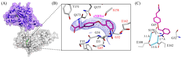
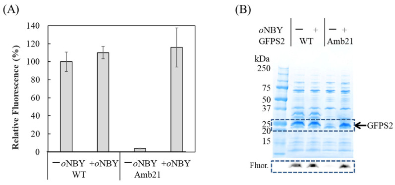
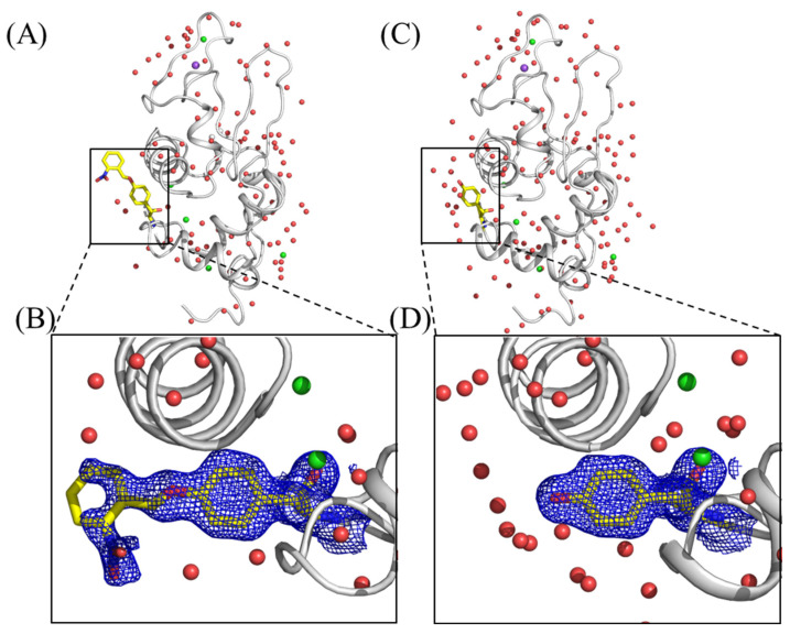
Genetically encoded caged amino acids can be used to control the dynamics of protein activities and cellular localization in response to external cues. In the present study, we revealed the structural basis for the recognition of O-(2-nitrobenzyl)-L-tyrosine (oNBTyr) by its specific variant of Methanocaldococcus jannaschii tyrosyl-tRNA synthetase (oNBTyrRS), and then demonstrated its potential availability for time-resolved X-ray crystallography. The substrate-bound crystal structure of oNBTyrRS at a 2.79 Å resolution indicated that the replacement of tyrosine and leucine at positions 32 and 65 by glycine (Tyr32Gly and Leu65Gly, respectively) and Asp158Ser created sufficient space for entry of the bulky substitute into the amino acid binding pocket, while Glu in place of Leu162 formed a hydrogen bond with the nitro moiety of oNBTyr. We also produced an oNBTyr-containing lysozyme through a cell-free protein synthesis system derived from the Escherichia coli B95. ΔA strain with the UAG codon reassigned to the nonnatural amino acid. Another crystallographic study of the caged protein showed that the site-specifically incorporated oNBTyr was degraded to tyrosine by light irradiation of the crystals. Thus, cell-free protein synthesis of caged proteins with oNBTyr could facilitate time-resolved structural analysis of proteins, including medically important membrane proteins.

Keywords: O-(2-nitrobenzyl)-L-tyrosine; X-ray crystallography; caged amino acid; cell-free protein synthesis; nonnatural amino acid.


Conflict of interest statement

The authors declare no conflict of interest.

Figures

Figure 1
Figure 1
The crystal structure of M. jannaschii oNBTyrRS (Y32G, L65G, F108E, D158S, L162E, and D286R) complexed with oNBTyr. (A) Dimeric structure of M. jannaschii oNBTyrRS bound to oNBTyr in the asymmetric unit. The substrate oNBTyr and water molecules are shown by green sticks and red spheres. (B) Close-up view of oNBTyr. The |2Fo − Fc| electron density map (contoured at 1.0 σ, blue mesh) corresponds to oNBTyr in the amino-acid binding site of oNBTyrRS. Mutated residues are indicated by the red letters. (C) Hydrogen-bonding interactions to recognize the o-nitrobenzyl group by oNBTyrRS. Numbers indicate the distance (Å) between two atoms connected by dashed lines.
Figure 2
Figure 2
Structural comparison of the substrate binding modes of various MjTyrRS. Structures of (A) oNBTyr in oNBTyrRS (this study); (B) tyrosine in the wild-type MjTyrRS (PDB: 1J1U [12]); (C) m-oNBDOPA in the corresponding variant of MjTyrRS (PDB: 5L7P [8]); (D) chemical structures of oNBTyr, oNPTyr, and m-oNB-DOPA.
Figure 3
Figure 3
Cell-free protein synthesis of N11-GFPS2 with (+) or without (−) o-nitrobenzyl Tyr (oNBY) supplementation using the cell extract from the strain B95.deltaA. Wild-type GFPS2 is indicated as “WT” and GFPS2 with Y21amber mutation is indicated as “Amb21”. (A) Fluorescent measurement of GFP in the reaction solutions. The excitation and emission wavelengths are 485 nm and 535 nm, respectively. Data represent the mean ± standard deviation (SD) of four independent experiments; (B) SDS-PAGE analysis of fractions purified by Ni-NTA resin. The 10–20% polyacrylamide gel was stained with CBB after electrophoresis. A portion of a fluorescent image of the gel framed by the dashed line is shown in the lower panel. The fluorescent imaging was performed by Vilber Bio Imaging FUSION FX with 470 nm and 535 nm filters. Molecular weights of protein standards (BIO-RAD Cat. No. 1610363) are indicated.
Figure 4
Figure 4
Crystal Structures of hen egg-white lysozyme (HEWL) with the incorporation of oNBTyr before (A) and after (C) photoactivation. Close-up views of the oNBTyr; (B) and decaged oNBTyr, Tyr; (D) at position 20 of HEWL are shown by stick yellow models. The |2Fo − Fc| electron density maps (0.6 σ) around Tyr20 are depicted by blue mesh. Water molecules, sodium, and chloride atoms are shown by red, purple, and light green spheres, respectively.

Similar articles

Cited by

References

    1. Engels J., Schlaeger E.J. Synthesis, structure, and reactivity of adenosine cyclic 3′,5′-phosphate benzyl triesters. J. Med. Chem. 1977;20:907–911. doi: 10.1021/jm00217a008. - DOI - PubMed
    1. Kaplan J.H., Forbush B., 3rd, Hoffman J.F. Rapid photolytic release of adenosine 5′-triphosphate from a protected analogue: Utilization by the Na: K pump of human red blood cell ghosts. Biochemistry. 1978;17:1929–1935. doi: 10.1021/bi00603a020. - DOI - PubMed
    1. Mayer G., Heckel A. Biologically active molecules with a “light switch”. Angew. Chem. Int. Ed. Engl. 2006;45:4900–4921. doi: 10.1002/anie.200600387. - DOI - PubMed
    1. Lee H.M., Larson D.R., Lawrence D.S. Illuminating the chemistry of life: Design, synthesis, and applications of “caged” and related photoresponsive compounds. ACS Chem. Biol. 2009;4:409–427. doi: 10.1021/cb900036s. - DOI - PMC - PubMed
    1. Gautier A., Gauron C., Volovitch M., Bensimon D., Jullien L., Vriz S. How to control proteins with light in living systems. Nat. Chem. Biol. 2014;10:533–541. doi: 10.1038/nchembio.1534. - DOI - PubMed

MeSH terms

LinkOut - more resources