

Cyto-Genotoxicity of Tritiated Stainless Steel and Cement Particles in Human Lung Cell Models
- PMID: 36142309
- PMCID: PMC9499181
- DOI: 10.3390/ijms231810398
Cyto-Genotoxicity of Tritiated Stainless Steel and Cement Particles in Human Lung Cell Models
Abstract
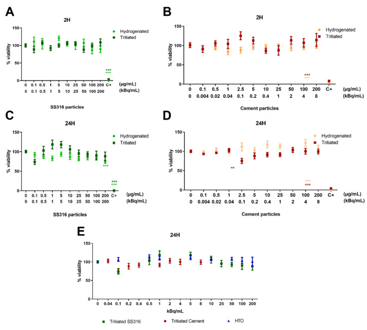
During the decommissioning of nuclear facilities, the tritiated materials must be removed. These operations generate tritiated steel and cement particles that could be accidentally inhaled by workers. Thus, the consequences of human exposure by inhalation to these particles in terms of radiotoxicology were investigated. Their cyto-genotoxicity was studied using two human lung models: the BEAS-2B cell line and the 3D MucilAirTM model. Exposures of the BEAS-2B cell line to particles (2 and 24 h) did not induce significant cytotoxicity. Nevertheless, DNA damage occurred upon exposure to tritiated and non-tritiated particles, as observed by alkaline comet assay. Tritiated particles only induced cytostasis; however, both induced a significant increase in centromere negative micronuclei. Particles were also assessed for their effects on epithelial integrity and metabolic activity using the MucilAirTM model in a 14-day kinetic mode. No effect was noted. Tritium transfer through the epithelium was observed without intracellular accumulation. Overall, tritiated and non-tritiated stainless steel and cement particles were associated with moderate toxicity. However, these particles induce DNA lesions and chromosome breakage to which tritium seems to contribute. These data should help in a better management of the risk related to the inhalation of these types of particles.
Keywords: BEAS-2B cells; DNA damage; MucilAirTM; cement particles; chromosome damage; cytotoxicity; in vitro testing; micronuclei; stainless steel particles; tritium.
Conflict of interest statement
The authors declare no conflict of interest. The funders had no role in the design of the study; in the collection, analyses, or interpretation of data; in the writing of the manuscript; or in the decision to publish the results.
Figures


Similar articles
-
Gaining insight into genotoxicity with the comet assay in inhomogenoeous exposure scenarios: The effects of tritiated steel and cement particles on human lung cells in an inhalation perspective.Toxicol In Vitro. 2023 Oct;92:105656. doi: 10.1016/j.tiv.2023.105656. Epub 2023 Jul 31. Toxicol In Vitro. 2023. PMID: 37532108
-
Bioaccumulation, release and genotoxicity of stainless steel particles in marine bivalve molluscs.Chemosphere. 2022 Sep;303(Pt 2):134914. doi: 10.1016/j.chemosphere.2022.134914. Epub 2022 May 16. Chemosphere. 2022. PMID: 35588874
-
Tritiated stainless steel (nano)particle release following a nuclear dismantling incident scenario: Significant exposure of freshwater ecosystem benthic zone.J Hazard Mater. 2024 Mar 5;465:133093. doi: 10.1016/j.jhazmat.2023.133093. Epub 2023 Nov 29. J Hazard Mater. 2024. PMID: 38056254
-
Genotoxicity of environmental agents assessed by the alkaline comet assay.Basic Clin Pharmacol Toxicol. 2005;96 Suppl 1:1-42. Basic Clin Pharmacol Toxicol. 2005. PMID: 15859009 Review.
-
The comet assay with multiple mouse organs: comparison of comet assay results and carcinogenicity with 208 chemicals selected from the IARC monographs and U.S. NTP Carcinogenicity Database.Crit Rev Toxicol. 2000 Nov;30(6):629-799. doi: 10.1080/10408440008951123. Crit Rev Toxicol. 2000. PMID: 11145306 Review.
References
-
- UNSCEAR . Sources, Effects and Risks of Ionizing Radiation. United Nations; New York, NY, USA: 2016.
-
- Matsumoto H., Shimada Y., Nakamura A.J., Usami N., Ojima M., Kakinuma S., Shimada M., Sunaoshi M., Hirayama R., Tauchi H. Health Effects Triggered by Tritium: How Do We Get Public Understanding Based on Scientifically Supported Evidence? J. Radiat. Res. 2021;62:557–563. doi: 10.1093/jrr/rrab029. - DOI - PMC - PubMed
-
- Liger K., Grisolia C., Cristescu I., Moreno C., Malard V., Coombs D., Markelj S. Overview of the TRANSAT (TRANSversal Actions for Tritium) Project. Nucl. Eng. Des. Fusion. 2018;136:168–172. doi: 10.1016/j.fusengdes.2018.01.037. - DOI
-
- Roch-Lefèvre S., Grégoire E., Martin-Bodiot C., Flegal M., Fréneau A., Blimkie M., Bannister L., Wyatt H., Barquinero J.-F., Roy L., et al. Cytogenetic Damage Analysis in Mice Chronically Exposed to Low-Dose Internal Tritium Beta-Particle Radiation. Oncotarget. 2018;9:27397–27411. doi: 10.18632/oncotarget.25282. - DOI - PMC - PubMed
MeSH terms
Substances
Grants and funding
LinkOut - more resources
Full Text Sources
