

Applications of Metabolomics in Calcium Metabolism Disorders in Humans
- PMID: 36142318
- PMCID: PMC9499180
- DOI: 10.3390/ijms231810407
Applications of Metabolomics in Calcium Metabolism Disorders in Humans
Abstract
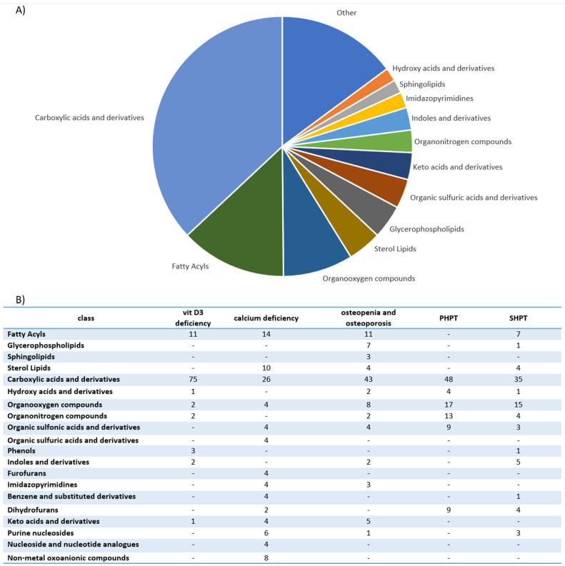
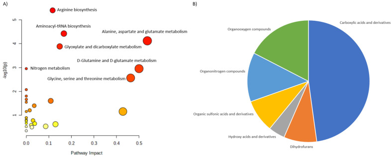
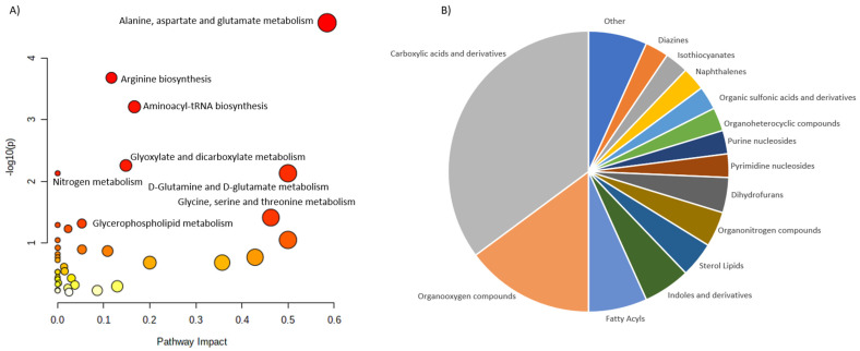
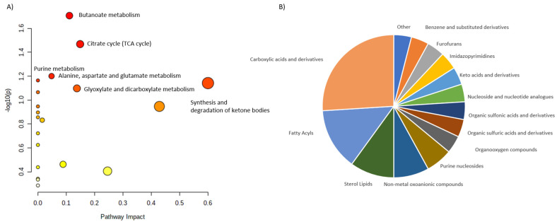
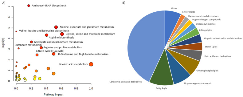
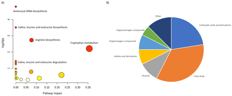
The pathogenesis of the disorders of calcium metabolism is not fully understood. This review discusses the studies in which metabolomics was applied in this area. Indeed, metabolomics could play an essential role in discovering biomarkers and elucidating pathological mechanisms. Despite the limited bibliography, the present review highlights the potential of metabolomics in identifying the biomarkers of some of the most common endocrine disorders, such as primary hyperparathyroidism (PHPT), secondary hyperparathyroidism (SHPT), calcium deficiency, osteoporosis and vitamin D supplementation. Metabolites related to above-mentioned diseorders were grouped into specific classes and mapped into metabolic pathways. Furthermore, disturbed metabolic pathways can open up new directions for the in-depth exploration of the basic mechanisms of these diseases at the molecular level.
Keywords: biomarkers; hypercalcemia; hypocalcemia; mass spectrometry; metabolites; metabolomics; parathyroid gland.
Conflict of interest statement
The authors declare no conflict of interest.
Figures
Similar articles
-
New parathyroid function index for the differentiation of primary and secondary hyperparathyroidism: a case-control study.BMC Endocr Disord. 2020 Jan 8;20(1):5. doi: 10.1186/s12902-019-0487-8. BMC Endocr Disord. 2020. PMID: 31914999 Free PMC article.
-
Vitamin D dependency: an inherited postnatal syndrome with secondary hyperparathyroidism.Pediatrics. 1970 Dec;46(6):871-80. Pediatrics. 1970. PMID: 5491441 No abstract available.
-
Secondary hyperparathyroidism.Mt Sinai J Med. 1973 May-Jun;40(3):462-73. Mt Sinai J Med. 1973. PMID: 4351493 No abstract available.
-
Cinacalcet HCl: a novel treatment for secondary hyperparathyroidism caused by chronic kidney disease.J Ren Nutr. 2006 Jul;16(3):253-8. doi: 10.1053/j.jrn.2006.04.010. J Ren Nutr. 2006. PMID: 16825031 Review.
-
Calcium Metabolic Disorders in Pregnancy: Primary Hyperparathyroidism, Pregnancy-Induced Osteoporosis, and Vitamin D Deficiency in Pregnancy.Endocrinol Metab Clin North Am. 2019 Sep;48(3):643-655. doi: 10.1016/j.ecl.2019.05.007. Epub 2019 Jun 14. Endocrinol Metab Clin North Am. 2019. PMID: 31345528 Review.
Cited by
-
Combining untargeted and targeted metabolomic profiling reveals principal differences between osteopenia, Osteoporosis and healthy controls.Aging Clin Exp Res. 2025 Jan 21;37(1):28. doi: 10.1007/s40520-024-02923-3. Aging Clin Exp Res. 2025. PMID: 39833609 Free PMC article.
-
New insight into primary hyperparathyroidism using untargeted metabolomics.Sci Rep. 2024 Sep 9;14(1):20987. doi: 10.1038/s41598-024-71423-1. Sci Rep. 2024. PMID: 39251672 Free PMC article.
-
Fighting Obesity-Related Micronutrient Deficiencies through Biofortification of Agri-Food Crops with Sustainable Fertilization Practices.Plants (Basel). 2022 Dec 12;11(24):3477. doi: 10.3390/plants11243477. Plants (Basel). 2022. PMID: 36559589 Free PMC article. Review.
-
Impact of subclinically hypocalcemic stress on the plasma metabolomic profile of dairy goats.Anim Biosci. 2025 May;38(5):981-992. doi: 10.5713/ab.24.0567. Epub 2025 Jan 24. Anim Biosci. 2025. PMID: 39901708 Free PMC article.
-
Effects of Menaquinone-7 on the Bone Health of Growing Rats under Calcium Restriction: New Insights from Microbiome-Metabolomics.Nutrients. 2023 Jul 31;15(15):3398. doi: 10.3390/nu15153398. Nutrients. 2023. PMID: 37571336 Free PMC article.
References
-
- Yu E., Sharma S. StatPearls. StatPearls Publishing; Treasure Island, FL, USA: 2022. Physiology, Calcium.
Publication types
MeSH terms
Substances
LinkOut - more resources
Full Text Sources
