

Exosomes Derived from Human Umbilical Cord Mesenchymal Stem Cells Accelerate Diabetic Wound Healing via Promoting M2 Macrophage Polarization, Angiogenesis, and Collagen Deposition
- PMID: 36142334
- PMCID: PMC9498995
- DOI: 10.3390/ijms231810421
Exosomes Derived from Human Umbilical Cord Mesenchymal Stem Cells Accelerate Diabetic Wound Healing via Promoting M2 Macrophage Polarization, Angiogenesis, and Collagen Deposition
Abstract
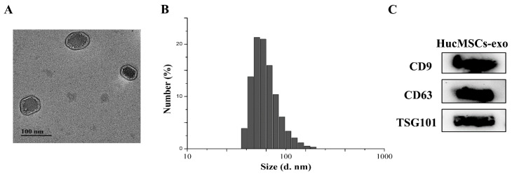
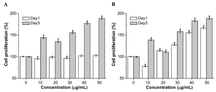
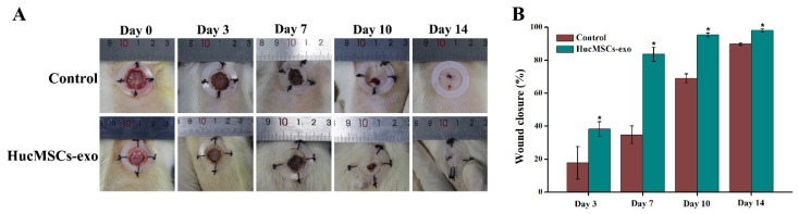
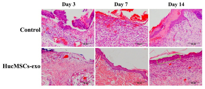
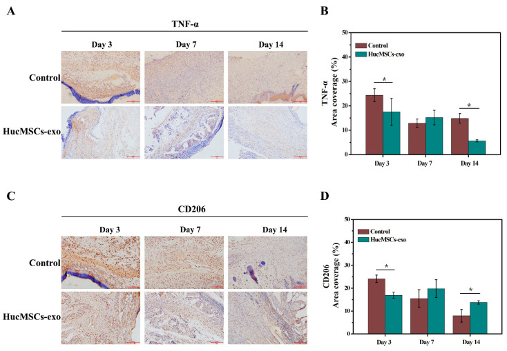
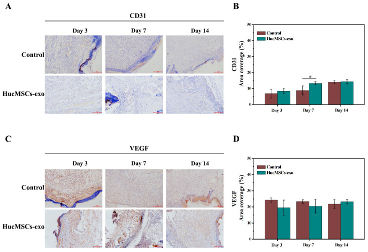
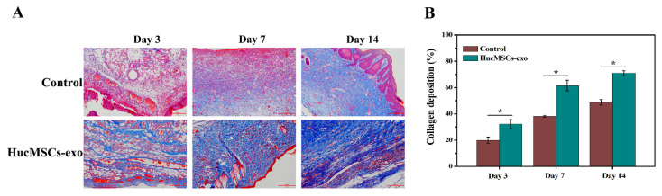
Some scholars have suggested that the clinical application of exosomes derived from human umbilical cord mesenchymal stem cells (hucMSCs-exo) might represent a novel strategy to improve diabetic wound healing. However, the mechanisms underlying the effects of hucMSCs-exo on wound healing remain poorly understood. This study aimed to identify the mechanism of hucMSCs-exo in treating diabetic wounds. HucMSCs-exo were isolated from human umbilical cord mesenchymal stem cells (hucMSCs) and subcutaneously injected into full-thickness wounds in diabetic rats. Wound healing closure rates and histological analysis were performed. The levels of tumor necrosis factor-α (TNF-α), macrophage mannose receptor (MMR/CD206), platelet endothelial cell adhesion molecule-1 (PECAM-1/CD31), and vascular endothelial growth factor (VEGF) were detected by immunohistochemistry. The degree of collagen deposition was examined using Masson's trichrome staining. Gross evaluation of wound healing was carried out from day 0 to 14 post-surgery, and the wound site was harvested for histology on days 3, 7, and 14 post-wounding. HucMSCs-exo transplantation increased diabetic wound healing. In vitro, hucMSCs-exo promoted the proliferation of human umbilical vein endothelial cells (HUVECs) and NIH-3T3 cells. In vivo, hucMSCs-exo reduced wound area and inflammatory infiltration and increased collagen fibers. In addition, wound tissues in the hucMSCs-exo group had higher CD206, CD31, and VEGF expressions and lower TNF-α levels than those in the control group on day 14. Our results demonstrated that hucMSCs-exo facilitated diabetic wound repair by inducing anti-inflammatory macrophages and promoting angiogenesis and collagen deposition.
Keywords: diabetes; exosomes; human-umbilical-cord-derived mesenchymal stem cells; macrophage polarization; wound healing.
Conflict of interest statement
The authors declare no conflict of interest.
Figures
Similar articles
-
Preparation of exosomes encapsulated nanohydrogel for accelerating wound healing of diabetic rats by promoting angiogenesis.Mater Sci Eng C Mater Biol Appl. 2021 Jan;120:111671. doi: 10.1016/j.msec.2020.111671. Epub 2020 Oct 22. Mater Sci Eng C Mater Biol Appl. 2021. PMID: 33545836
-
MSC Exosomes Containing Valproic Acid Promote Wound Healing by Modulating Inflammation and Angiogenesis.Molecules. 2024 Sep 9;29(17):4281. doi: 10.3390/molecules29174281. Molecules. 2024. PMID: 39275128 Free PMC article.
-
Collagen sponge scaffolds loaded with Trichostatin A pretreated BMSCs-derived exosomes regulate macrophage polarization to promote skin wound healing.Int J Biol Macromol. 2024 Jun;269(Pt 2):131948. doi: 10.1016/j.ijbiomac.2024.131948. Epub 2024 Apr 28. Int J Biol Macromol. 2024. PMID: 38688338
-
The Therapeutic Potential of Human Umbilical Cord Mesenchymal Stromal Cells Derived Exosomes for Wound Healing: Harnessing Exosomes as a Cell-free Therapy.J Stem Cells Regen Med. 2024 May 31;20(1):14-23. doi: 10.46582/jsrm.2003003. eCollection 2024. J Stem Cells Regen Med. 2024. PMID: 39044811 Free PMC article. Review.
-
Exosomes from mesenchymal stem cells: Potential applications in wound healing.Life Sci. 2024 Nov 15;357:123066. doi: 10.1016/j.lfs.2024.123066. Epub 2024 Sep 19. Life Sci. 2024. PMID: 39306326 Review.
Cited by
-
Exosomal peptides and proteins in wound healing and skin regeneration.Naunyn Schmiedebergs Arch Pharmacol. 2025 Jul 8. doi: 10.1007/s00210-025-04426-y. Online ahead of print. Naunyn Schmiedebergs Arch Pharmacol. 2025. PMID: 40627201 Review.
-
Exosome-Based Therapies in Dermatology.Aesthetic Plast Surg. 2025 Aug 8. doi: 10.1007/s00266-025-05073-7. Online ahead of print. Aesthetic Plast Surg. 2025. PMID: 40779041 Review.
-
Emerging Nanotherapeutic Approaches for Diabetic Wound Healing.Int J Nanomedicine. 2024 Aug 27;19:8815-8830. doi: 10.2147/IJN.S476006. eCollection 2024. Int J Nanomedicine. 2024. PMID: 39220193 Free PMC article. Review.
-
Exosomes Derived from Human Amniotic Mesenchymal Stem Cells Facilitate Diabetic Wound Healing by Angiogenesis and Enrich Multiple lncRNAs.Tissue Eng Regen Med. 2023 Apr;20(2):295-308. doi: 10.1007/s13770-022-00513-w. Epub 2023 Jan 25. Tissue Eng Regen Med. 2023. PMID: 36696086 Free PMC article.
-
Understanding molecular characteristics of extracellular vesicles derived from different types of mesenchymal stem cells for therapeutic translation.Extracell Vesicle. 2024 Jun;3:100034. doi: 10.1016/j.vesic.2024.100034. Epub 2024 Mar 2. Extracell Vesicle. 2024. PMID: 38957857 Free PMC article.
References
-
- Diabetes Branch of Chinese Medical Association Guideline for The Prevention and Treatment of Type 2 Diabetes Mellitus in China (2020 edition) Zhonghua Tang Niao Bing Za Zhi. 2021;13:315–409.
MeSH terms
Substances
LinkOut - more resources
Full Text Sources
