. 2022 Sep 10;23(18):10498.
doi: 10.3390/ijms231810498.

Identification of Adipose Tissue as a Reservoir of Macrophages after Acute Myocardial Infarction

Affiliations

Identification of Adipose Tissue as a Reservoir of Macrophages after Acute Myocardial Infarction

Ingrid Gomez et al. Int J Mol Sci. .

Abstract

Medullary and extra-medullary hematopoiesis has been shown to govern inflammatory cell infiltration and subsequently cardiac remodeling and function after acute myocardial infarction (MI). Emerging evidence positions adipose tissue (AT) as an alternative source of immune cell production. We, therefore, hypothesized that AT could act as a reservoir of inflammatory cells that participate in cardiac homeostasis after MI. To reveal the distinct role of inflammatory cells derived from AT or bone marrow (BM), chimeric mice were generated using standard repopulation assays. We showed that AMI increased the number of AT-derived macrophages in the cardiac tissue. These macrophages exhibit pro-inflammatory characteristics and their specific depletion improved cardiac function as well as decreased infarct size and interstitial fibrosis. We then reasoned that the alteration of AT-immune compartment in type 2 diabetes could, thus, contribute to defects in cardiac remodeling. However, in these conditions, myeloid cells recruited in the infarcted heart mainly originate from the BM, and AT was no longer used as a myeloid cell reservoir. Altogether, we showed here that a subpopulation of cardiac inflammatory macrophages emerges from myeloid cells of AT origin and plays a detrimental role in cardiac remodeling and function after MI. Diabetes abrogates the ability of AT-derived myeloid cells to populate the infarcted heart.

Keywords: adipose tissue; diabetes; macrophages; myocardial infarction.


Conflict of interest statement

The authors declare no conflict of interest.

Figures

Figure 1
Figure 1
Establishment of chimeric mice models to reveal the distinct role of adipose tissue or bone marrow-derived inflammatory cells. (A) Hematopoietic chimera strategy: 2.103 hematopoietic stem cells (HSC) sorted from sub-cutaneous adipose tissue (AT) or bone marrow (BM) of mTmG mice were co-injected with 2.105 total BM cells from C57Bl6 mice into lethally irradiated C57Bl6 recipients. Eight weeks after hematopoietic reconstitution, chimeric mice were subjected to myocardial infarction. (B) Representative dot plots of flow cytometry analyses showing the chimerism (tdT+ cells among CD45+ cells; open histogram) or wild type cells (dT-; grey histogram) in the AT of AT- chimeric mice (n = 4). (C) Quantification of the chimerism in sub-cutaneous AT, BM, heart and spleen of AT- and BM-chimeric mice (n = 4–6). (D) Ejection Fraction in unoperated AT- and BM chimeric mice (n = 8). (E) Quantification of ejection fraction, infarct size and interstitial fibrosis in sham and infarcted chimeric mice (n = 5). Comparisons between 2 group were performed using Dunn’s multiple comparisons test. ** p < 0.01.
Figure 2
Figure 2
Adipose tissue is a reservoir of monocytes and macrophages populating the infarcted heart. (A) Quantification of CD45+/dT+ in blood and heart of sham and infarcted AT-chimeric mice (n = 4–9). (B) Percentage of dT+ cells in myeloid and lymphoid populations, in the heart of AT-mice, 7 days post MI (n = 7). (C) Quantification of dT+ and dT- Ly6Chigh, Ly6Clow monocytes and macrophages in the heart, 7 days after MI in BM- and AT-chimeric mice (n = 4–10). In the stacked bars, black columns indicate the number of dT- cells and colored columns the number of dT+ cells in BM-mice (grey) or AT-mice (blue). (D) Quantification of IL-1α, IL-6 and IL-10 production in cardiac macrophages sorted from BM- and AT-mice 7 days post MI (n = 4–6). Comparisons between groups were performed using Dunn’s multiple comparisons test (B) or Mann-Whitney Test (D). * p < 0.05; ** p < 0.01.
Figure 3
Figure 3
Depletion of adipose tissue-derived CD11c+ macrophages improves cardiac remodeling and function after acute MI. (A) Schematic representation of experimental procedure: lethally irradiated mT/mG recipient mice were co-injected with hematopoietic stem cells (HSC) sorted from sub-cutaneous adipose tissue (AT) or bone marrow (BM) and total BM cells isolated from CD11c-DTR and mT/mG donor mice, respectively. Eight weeks after reconstitution, chimeric mice were treated with diphtheria toxin (DT) or PBS as control at day 0, 3 and 6 after the ischemic insult. (B) Quantification of CD11c+ cells and CD11c+ macrophages in the heart, 7 days post-MI in BM- and AT-mice (n = 3–4). (C) Representative photomicrographs and quantification of ejection fraction, infarction size (scale bar, 2 mm), interstitial fibrosis (scale bar, 10 µm) and capillary density (scale bar, 10µm) in BM and AT-mice treated with or without DT (n = 4–8). Comparisons between groups were performed using Dunn’s multiple comparisons test. * p < 0.05 in DT vs. PBS treated mice.
Figure 4
Figure 4
Depletion of adipose tissue-derived CD11c+ macrophages lowers cardiac inflammation. IL-1α, IL-6, IFNγ and TNFα levels in the heart (A) and the blood (B) of AT- and BM- chimeric mice treated with or without diphtheria toxin (DT), 7 days post- MI (n = 4–6). Comparisons between groups were performed using Dunn’s multiple comparisons test. * p < 0.05 in DT vs. PBS treated mice.
Figure 5
Figure 5
High Fat Diet worsens cardiac remodeling and function. (A) Schematic representation of experimental procedure: lethally irradiated recipient mice were co-injected with hematopoietic stem cells (HSC) sorted from sub-cutaneous adipose tissue (AT) or bone marrow (BM) and total BM cells isolated from mT/mG donor and C57Bl6 donor mice, respectively. Eight weeks after reconstitution, mice were then fed a normal Chow (NC) or a high fat diet (HFD) for 2 months. (B) Ejection fraction and (C) infarct size as well as interstitial fibrosis in BM- and AT- mice fed a normal Chow (NC) or a high fat diet (HFD) for 2 months and then challenged with MI (n = 4–10). Comparisons between groups were performed using Dunn’s multiple comparisons test. * p < 0.05 in HFD vs. NC fed mice.
Figure 6
Figure 6
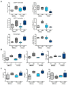
Diabetic chimeric mice exhibit higher cardiac inflammation. (A) Quantification of total and dT+ Ly6CHighand Ly6CLow monocytes, and macrophages in the heart of BM- and AT-mice fed a normal chow (NC) or a high fat diet (HFD) for 8 weeks (n = 4–8). (B,C) Cytokine levels in the heart (B) and the blood (C) of BM- and AT-mice fed a normal chow (NC) or a high fat diet (HFD) for 8 weeks (n = 4–5). Analyses have been performed 7 days after the onset of acute MI. Comparisons between groups were performed using Dunn’s multiple comparisons test. * p < 0.05 and ** p < 0.01 in HFD vs. NC fed mice.
Figure 7
Figure 7
Adipose tissue-derived CD11c+ macrophage depletion did not regulate cardiac function and remodeling in diabetic mice. (A) Ejection fraction, interstitial fibrosis, infarct size and capillary density in diabetic BM- and AT-chimeric mice treated with or without diphtheria toxin (DT) (n = 6–8). (B) Monocyte and macrophage number in the heart of diabetic BM- and AT-chimeric mice treated with or without DT, (n = 6–8). (C) IL-1α, IL-6 and IFNγ levels in the heart of diabetic BM- and AT-chimeric mice treated with or without diphtheria toxin (DT) (n = 4–6). Comparisons between groups were performed using Dunn’s multiple comparisons test (* p < 0.05 in DT vs. PBS treated mice).

References

    1. Han J., Koh Y.J., Moon H.R., Ryoo H.G., Cho C.H., Kim I., Koh G.Y. Adipose tissue is an extramedullary reservoir for functional hematopoietic stem and progenitor cells. Blood. 2010;115:957–964. doi: 10.1182/blood-2009-05-219923. - DOI - PubMed
    1. Poglio S., De Toni F., Lewandowski D., Minot A., Arnaud E., Barroca V., Laharrague P., Casteilla L., Cousin B. In situ production of innate immune cells in murine white adipose tissue. Blood. 2012;120:4952–4962. doi: 10.1182/blood-2012-01-406959. - DOI - PubMed
    1. Cousin B., Casteilla L., Laharrague P., Luche E., Lorsignol A., Cuminetti V., Paupert J. Immuno-metabolism and adipose tissue: The key role of hematopoietic stem cells. Biochimie. 2016;124:21–26. doi: 10.1016/j.biochi.2015.06.012. - DOI - PubMed
    1. Rabiller L., Robert V., Arlat A., Labit E., Ousset M., Salon M., Coste A., Costa-Fernandes D., Monsarrat P., Ségui B., et al. Driving regeneration, instead of healing, in adult mammals: The decisive role of resident macrophages through efferocytosis. NPJ Regen. Med. 2021;6:1–12. doi: 10.1038/s41536-021-00151-1. - DOI - PMC - PubMed
    1. Poglio S., De Toni-Costes F., Arnaud E., Laharrague P., Espinosa E., Casteilla L., Cousin B. Adipose tissue as a dedicated reservoir of functional mast cell progenitors. Stem Cells. 2010;28:2065–2072. doi: 10.1002/stem.523. - DOI - PubMed

LinkOut - more resources