Review
. 2022 Sep 10;23(18):10490.
doi: 10.3390/ijms231810490.

Interaction between Alzheimer's Disease and Cerebral Small Vessel Disease: A Review Focused on Neuroimaging Markers

Affiliations
Review

Interaction between Alzheimer's Disease and Cerebral Small Vessel Disease: A Review Focused on Neuroimaging Markers

Si Eun Kim et al. Int J Mol Sci. .

Abstract

Alzheimer's disease (AD) is characterized by the presence of β-amyloid (Aβ) and tau, and subcortical vascular cognitive impairment (SVCI) is characterized by cerebral small vessel disease (CSVD). They are the most common causes of cognitive impairment in the elderly population. Concurrent CSVD burden is more commonly observed in AD-type dementia than in other neurodegenerative diseases. Recent developments in Aβ and tau positron emission tomography (PET) have enabled the investigation of the relationship between AD biomarkers and CSVD in vivo. In this review, we focus on the interaction between AD and CSVD markers and the clinical effects of these two markers based on molecular imaging studies. First, we cover the frequency of AD imaging markers, including Aβ and tau, in patients with SVCI. Second, we discuss the relationship between AD and CSVD markers and the potential distinct pathobiology of AD markers in SVCI compared to AD-type dementia. Next, we discuss the clinical effects of AD and CSVD markers in SVCI, and hemorrhagic markers in cerebral amyloid angiopathy. Finally, this review provides both the current challenges and future perspectives for SVCI.

Keywords: Alzheimer’s disease; cerebral small vessel disease; interaction; positron emission tomography; subcortical vascular cognitive impairment; tau; ß-Amyloid.


Conflict of interest statement

The authors declare no conflict of interest.

Figures
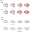
Figure 1
Figure 1
Spreading order of Aβ and tau in SVCI and AD, respectively. (a) Spreading pattern of Aβ in SVCI (a-1) and AD (a-2); (b) Spreading pattern of tau in SVCI (b-1) and AD (b-2). In the order of spreading Aβ in SVCI, unlike AD, Aβ accumulates in the occipital area before the temporal and frontal regions. In contrast to AD, tau accumulates in the fusiform gyrus and inferior temporal gyrus before the parahippocampal cortex in SVCI patients. Aβ—β-amyloid; SVCI—subcortical vascular cognitive impairment; AD—Alzheimer’s disease.
Figure 2
Figure 2
Potential mechanism of Aβ and tau deposition in SVCI. The CSVD burden is associated with Aβ and tau deposition in SVCI. Ischemic events can lead to Aβ deposition by reducing Aβ clearance via BBB breakdown or deficits in perivascular drainage of Aβ from the brain interstitial fluid. BBB breakdown causes faulty transport of Aβ by reducing LRP1 levels and increasing RAGE levels, resulting in impaired clearance of toxic Aβ species. Aβ accelerates the tau hyperphosphorylation by mediating the activation of protein kinases, including CDK-5 and GSK-3β. In addition, Aβ induces the activation of caspase-3 and calpain-1, and the cleavage of tau generates neurotoxic tau fragments. The association between Aβ and tau aggregation may involve microglial activation. Soluble Aβ oligomers have been known to activate microglial cells. Microglial activation precedes tau aggregation and facilitates tau hyperphosphorylation through cytokine release and the subsequent NFT formation. Vascular risk factors can also induce tau accumulation. Ischemia caused by vascular injury may activate CDK-5 and GSK-3 β, resulting in tau phosphorylation. Moreover, vascular risk factors and the accumulation of Aβ plaques lead to oxidative stress. Oxidative stress may also be caused by several mechanisms, such as mitochondrial dysfunction or inflammatory responses. It may manifest as damage to synapses and changes in Ca2+ homeostasis, resulting in an apoptotic cascade and neurotoxicity. CSVD—cerebral small vessel disease; Aβ—β-amyloid; BBB—blood–brain barrier; LRP1—low-density lipoprotein receptor-related protein 1; RAGE—receptor for advanced glycosylation end products; CDK-5—cyclin-dependent kinase 5; GSK-3β—glycogen synthase kinase 3β; NFTs—neurofibrillary tangles; ISF—interstitial fluid; CSF—cerebrospinal fluid; WMH—white matter hyperintensity; BM—basement membrane.
Figure 3
Figure 3
Mechanisms of SVCI. SVCI is caused by various factors, including increasing age, diabetes mellitus, hypertension, genetic predisposition such as APOE ε4 and ε2, and arteriosclerosis. This can lead to Aβ deposition by impairing Aβ clearance. SVCI is also related to widespread white matter hyperintensities or multiple lacunar infarctions, which have been gradually deposited in subcortical regions for several decades. CAA is another factor known to be associated with SVCI. SVCI—subcortical vascular cognitive impairment; APOE—apolipoprotein E; Aβ—β-amyloid; BBB—blood–brain barrier; CAA—cerebral amyloid angiopathy.

Similar articles

Cited by

References

    1. Scott K.R., Barrett A.M. Dementia syndromes: Evaluation and treatment. Expert Rev. Neurother. 2007;7:407–422. doi: 10.1586/14737175.7.4.407. - DOI - PMC - PubMed
    1. Roman G.C. Vascular dementia: Distinguishing characteristics, treatment, and prevention. J. Am. Geriatr. Soc. 2003;51((Suppl. S5)):S296–S304. doi: 10.1046/j.1532-5415.5155.x. - DOI - PubMed
    1. Wenk G.L. Neuropathologic changes in Alzheimer’s disease. J. Clin. Psychiatry. 2003;64((Suppl. 9)):7–10. - PubMed
    1. Jack C.R., Jr., Bennett D.A., Blennow K., Carrillo M.C., Dunn B., Haeberlein S.B., Holtzman D.M., Jagust W., Jessen F., Karlawish J., et al. NIA-AA Research Framework: Toward a biological definition of Alzheimer’s disease. Alzheimer’s Dement. J. Alzheimer’s Assoc. 2018;14:535–562. doi: 10.1016/j.jalz.2018.02.018. - DOI - PMC - PubMed
    1. Jack C.R., Jr., Wiste H.J., Therneau T.M., Weigand S.D., Knopman D.S., Mielke M.M., Lowe V.J., Vemuri P., Machulda M.M., Schwarz C.G., et al. Associations of Amyloid, Tau, and Neurodegeneration Biomarker Profiles With Rates of Memory Decline Among Individuals Without Dementia. Jama. 2019;321:2316–2325. doi: 10.1001/jama.2019.7437. - DOI - PMC - PubMed

MeSH terms