. 2022 Sep 10;23(18):10510.
doi: 10.3390/ijms231810510.

Thebromine Targets Adenosine Receptors to Control Hippocampal Neuronal Function and Damage

Affiliations

Thebromine Targets Adenosine Receptors to Control Hippocampal Neuronal Function and Damage

Pedro Valada et al. Int J Mol Sci. .

Abstract

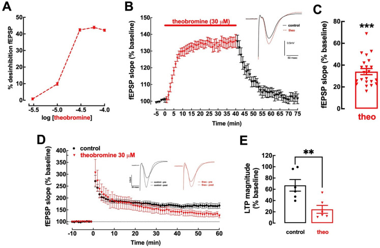
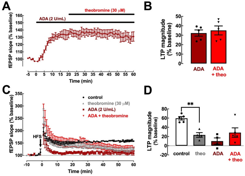
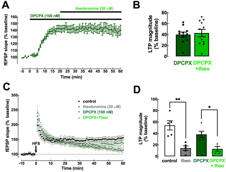
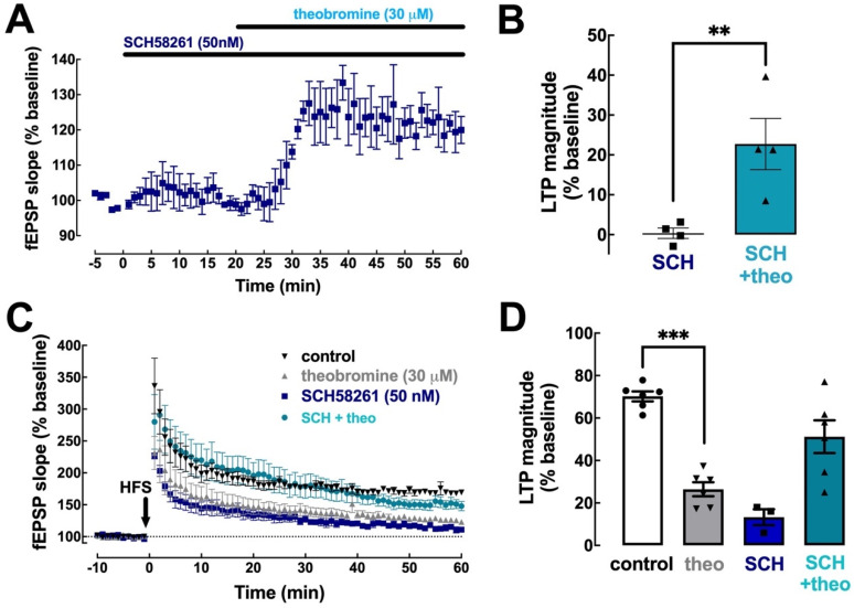
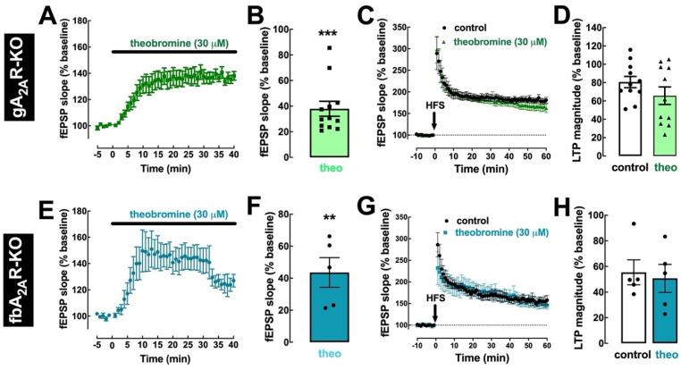
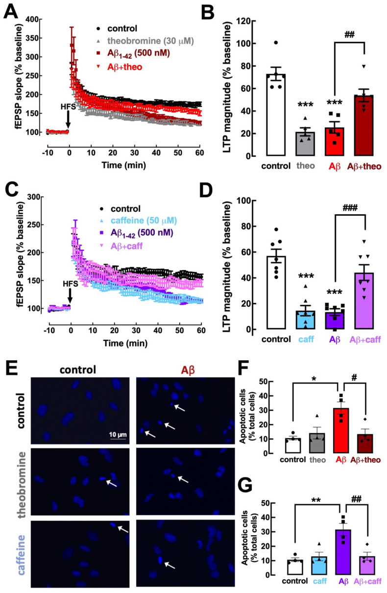
Theobromine is a caffeine metabolite most abundant in dark chocolate, of which consumption is linked with a lower risk of cognitive decline. However, the mechanisms through which theobromine affects neuronal function remain ill-defined. Using electrophysiological recordings in mouse hippocampal synapses, we now characterized the impact of a realistic concentration of theobromine on synaptic transmission and plasticity. Theobromine (30 μM) facilitated synaptic transmission while decreasing the magnitude of long-term potentiation (LTP), with both effects being blunted by adenosine deaminase (2 U/mL). The pharmacological blockade of A1R with DPCPX (100 nM) eliminated the theobromine-dependent facilitation of synaptic transmission, whereas the A2AR antagonist SCH58261 (50 nM), as well as the genetic deletion of A2AR, abrogated the theobromine-induced impairment of LTP. Furthermore, theobromine prevented LTP deficits and neuronal loss, respectively, in mouse hippocampal slices and neuronal cultures exposed to Aβ1-42 peptides, considered a culprit of Alzheimer's disease. Overall, these results indicate that theobromine affects information flow via the antagonism of adenosine receptors, normalizing synaptic plasticity and affording neuroprotection in dementia-related conditions in a manner similar to caffeine.

Keywords: Alzheimer’s disease; adenosine receptors; caffeine; synaptic plasticity; synaptic transmission; theobromine.


Conflict of interest statement

R.A.C. is a scientific advisor of the Institute for Scientific Information on Coffee (ISIC). All other authors declare no competing financial interests.

Figures

Figure 1
Figure 1
Theobromine alters basal synaptic transmission and synaptic plasticity. Theobromine increased basal synaptic transmission in Schaffer fibers-CA1 pyramid synapses of mouse hippocampal slices in a concentration-dependent manner, reaching a plateau after 30 μM (A). Theobromine (theo, 30 μM) rapidly and reversibly increased basal synaptic transmission (B,C), and the insert in (B) displays representative field extracellular postsynaptic potential (fEPSP) recorded in ACSF medium (control conditions, black filled line) and in the presence of theobromine (red dashed line). Additionally, theobromine significantly decreased the magnitude of long-term potentiation (LTP): the time course of fEPSP recordings (D) shows a sustained increase in fEPSP slope after delivering a high-frequency stimulation train (HFS: one train of 100 pulses of 1 Hz for 1 s), which is larger in ACSF (black symbols) than in the presence of 30 μM theobromine (red symbols), as quantified in (E). The inserts in (D) show two superimposed pairs fEPSP collected before (filled traces) and 60 min after HFS (dashed lines) in control conditions (black traces on the left) and in the presence of theobromine (red traces on the right). Data are mean ± S.E.M. of 5–22 (A), 22 (B,C) or 6 (D,E) experiments. The asterisks denote significant differences: ** p < 0.01 vs. control, Student’s t test (E); *** p < 0.001 vs. baseline, one-sample t test (C).
Figure 2
Figure 2
Clearance of endogenous extracellular adenosine abolishes the effects of theobromine on synaptic transmission and plasticity. Adenosine deaminase (ADA, 2 U/mL) enhanced hippocampal basal synaptic transmission (A,B) and abrogates the effects of theobromine (30 μM). ADA (2 U/mL) also decreased the magnitude of long-term potentiation (C,D) induced by a high-frequency stimulation train (HFS: one train of 100 pulses of 1 Hz for 1 s) in Schaffer fiber-CA1 pyramid synapses of mouse hippocampal slices and abrogates the effects of theobromine (theo, 30 μM). This shows that these measured effects of theobromine are strictly dependent on the presence of endogenous extracellular adenosine. Data are mean ± S.E.M. of 5 (A,B) and 4–5 (C,D) experiments. The asterisks denote significant differences: ** p < 0.01 vs. control, two-way ANOVA with Tukey’s post hoc test.
Figure 3
Figure 3
Blockade of A1R eliminates the impact of theobromine on basal synaptic transmission but not on synaptic plasticity. The selective A1R antagonist DPCPX (100 nM) enhanced basal synaptic transmission and occluded the effect of theobromine (theo, 30 μM) on synaptic transmission (A,B). In contrast, DPCPX did not modify the magnitude of long-term potentiation (LTP), induced with a high-frequency stimulation train (HFS: one train of 100 pulses of 1 Hz for 1 s), recorded in Schaffer fiber-CA1 pyramid synapses of mouse hippocampal slices and failed to modify the effects of theobromine on LTP magnitude (C,D). Data are mean ± S.E.M. of 11 (A,B) and 5–6 (C,D) experiments. The asterisks denote significant differences: * p < 0.05 and ** p < 0.01, two-way ANOVA with Tukey’s post hoc test.
Figure 4
Figure 4
Blockade of A2AR prevents the effect of theobromine on long-term potentiation. The selective A2AR antagonist SCH58261 (SCH, 50 nM) did not modify basal synaptic transmission nor altered the effect of theobromine (theo, 30 μM) on basal synaptic transmission in mouse hippocampal slices (A,B). In the presence of SCH58261 (50 nM), theobromine (30 μM) failed to decrease LTP magnitude (C,D), induced with a high-frequency stimulation train (HFS: one train of 100 pulses of 1 Hz for 1 s) in Schaffer fiber-CA1 pyramid synapses of mouse hippocampal slices. Data are mean ± S.E.M. of 4 (A,B) and 3–6 (C,D) experiments. The asterisks denote significant differences: ** p < 0.05 vs. SCH58261, Student’s t test (B); *** p < 0.001 vs. control, two-way ANOVA with Tukey’s post hoc test.
Figure 5
Figure 5
Genetic deletion of A2AR prevents the effect of theobromine on long-term potentiation. In slices from knockout mice where A2AR were deleted globally (gA2AR-KO, (AD)) or selectively in forebrain neurons (fbA2AR-KO, (EH)), theobromine (theo, 30 μM) still enhanced basal synaptic transmission (A,B,E,F), but did not modify the magnitude of LTP (C,D,G,H), induced with a high-frequency stimulation train (HFS: one train of 100 pulses of 1 Hz for 1 s) in Schaffer fiber-CA1 pyramid synapses of mouse hippocampal slices. Data are mean ± S.E.M. of 12 (A,B), 11 (C,D), and 5 (EH) experiments. The asterisks denote significant differences: ** p < 0.01, *** p < 0.001 vs. baseline, one-sample t test.
Figure 6
Figure 6
Theobromine and caffeine prevent synaptic plasticity deficits and neurotoxicity induced by Aβ1–42 modelling early Alzheimer’s disease. The superfusion of the mouse hippocampal slices with Aβ1–42 (50 nM) during 40 min induced a significant decrease in LTP magnitude (AD) that was prevented by the presence of either theobromine (theo, 30 μM) (A,B) or caffeine (caff, 50 μM) (C,D). Likewise, primary neuronal cultures treated with Aβ1–42 (500 nM) during 24 h exhibit a significant increase in the number of apoptotic neurons (EG), which were identified as cells with condensed nuclei with an irregular form often fragmented, displaying a more intense light blue staining with DAPI, as indicated by the arrows in the photographs (E). This neurotoxicity of Aβ1–42 was prevented both by theobromine (30 μM) (E,G) or caffeine (50 μM) (F,G). Data are mean ± S.E.M. of 5–6 (A,B), 7 (C,D), and 4 (EG) experiments, where the counting of apoptotic-like neurons versus the total number of neurons was calculated in 4 field per coverslip. The scale bar (10 μm) of the first photograph in (E) applies to all other photographs. The asterisks denote significant differences: * p < 0.05, ** p < 0.01, *** p < 0.001 vs. control, two-way ANOVA with Tukey’s post hoc test; # p < 0.05, ## p < 0.01, ### p < 0.001 vs. Aβ1–42, two-way ANOVA with Tukey’s post hoc test.

Similar articles

Cited by

References

    1. Smith A. Effects of caffeine on human behavior. Food Chem. Toxicol. 2002;40:1243–1255. doi: 10.1016/S0278-6915(02)00096-0. - DOI - PubMed
    1. Angelucci M.E.M., Cesário C., Hiroi R.H., Rosalen P.L., Da Cunha C. Effects of caffeine on learning and memory in rats tested in the Morris water maze. Braz. J. Med. Biol. Res. 2002;35:1201–1208. doi: 10.1590/S0100-879X2002001000013. - DOI - PubMed
    1. Borota D., Murray E., Keceli G., Chang A., Watabe J.M., Ly M., Toscano J.P., Yassa M.A. Post-study caffeine administration enhances memory consolidation in humans. Nat. Neurosci. 2014;17:201–203. doi: 10.1038/nn.3623. - DOI - PMC - PubMed
    1. Dong X., Li S., Sun J., Li Y., Zhang D. Association of coffee, decaffeinated coffee and caffeine intake from coffee with cognitive performance in older adults: National Health and Nutrition Examination Survey (NHANES) 2011–2014. Nutrients. 2020;12:840. doi: 10.3390/nu12030840. - DOI - PMC - PubMed
    1. Eskelinen M.H., Kivipelto M. Caffeine as a protective factor in dementia and Alzheimer’s disease. J. Alzheimers Dis. 2010;20((Suppl. S1)):S167–S174. doi: 10.3233/JAD-2010-1404. - DOI - PubMed