

Flow Cytometric Identification of Hematopoietic and Leukemic Blast Cells for Tailored Clinical Follow-Up of Acute Myeloid Leukemia
- PMID: 36142442
- PMCID: PMC9506284
- DOI: 10.3390/ijms231810529
Flow Cytometric Identification of Hematopoietic and Leukemic Blast Cells for Tailored Clinical Follow-Up of Acute Myeloid Leukemia
Abstract
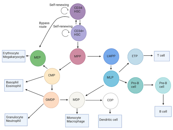
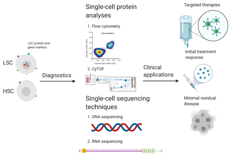
Acute myeloid leukemia (AML) is a myeloid malignancy that is characterized by the accumulation of leukemic blast cells, which originate from hematopoietic stem cells that have undergone leukemic transformation and/or are more mature progenitors that have gained stemness features. Currently, no consensus exists for the flow cytometric identification of normal blast cells and their leukemic counterparts by their antigenic expression profile. Differentiating between the benign cells and the malignant cells is crucial for the further deployment of immunophenotype panels for the clinical follow-up of AML patients. This review provides an overview of immunophenotypic markers that allow the identification of leukemic blast cells in the bone marrow with multiparameter flow cytometry. This technique allows the identification of hematopoietic blast cells at the level of maturing cells by their antigen expression profile. While aberrant antigen expression of a single immunophenotypic marker cell cannot be utilized in order to differentiate leukemic blast cells from normal blast cells, combinations of multiple immunophenotypic markers can enable the distinction of normal and leukemic blast cells. The identification of these markers has provided new perspectives for tailored clinical follow-up, including therapy management, diagnostics, and prognostic purposes. The immunophenotypic marker panels, however, should be developed by carefully considering the variable antigen marker expression profile of individual patients.
Keywords: CD marker expression profiles; distinction; flow cytometry; hematopoietic stem cells; leukemic stem cells.
Conflict of interest statement
The authors declare no conflict of interest.
Figures
Similar articles
-
Immunophenotypic features of leukemic stem cells and bulk of blasts in acute myeloid leukemia.Exp Oncol. 2019 Sep;41(3):207-209. doi: 10.32471/exp-oncology.2312-8852.vol-41-no-3.13492. Exp Oncol. 2019. PMID: 31569935
-
Prediction of relapse of pediatric acute myeloid leukemia by use of multidimensional flow cytometry.J Natl Cancer Inst. 1996 Oct 16;88(20):1483-8. doi: 10.1093/jnci/88.20.1483. J Natl Cancer Inst. 1996. PMID: 8841024
-
Clinical impact of leukemic blast heterogeneity at diagnosis in cytogenetic intermediate-risk acute myeloid leukemia.Cytometry B Clin Cytom. 2012 May;82(3):123-31. doi: 10.1002/cyto.b.20633. Epub 2012 Feb 10. Cytometry B Clin Cytom. 2012. PMID: 22328535
-
Immunophenotype characterization of hematopoietic stem cells, progenitor cells restricted to myeloid lineage and their leukemia counterparts.Neoplasma. 2010;57(5):392-400. doi: 10.4149/neo_2010_05_392. Neoplasma. 2010. PMID: 20568892 Review.
-
Study of morphocytochemical and immunophenotypic features of acute leukemia stem cells.Exp Oncol. 2008 Jun;30(2):102-5. Exp Oncol. 2008. PMID: 18566571 Review.
Cited by
-
Phenotypic Analysis of Hematopoietic Stem and Progenitor Cell Populations in Acute Myeloid Leukemia Based on Spectral Flow Cytometry, a 20-Color Panel, and Unsupervised Learning Algorithms.Int J Mol Sci. 2024 Feb 29;25(5):2847. doi: 10.3390/ijms25052847. Int J Mol Sci. 2024. PMID: 38474094 Free PMC article.
-
Epigenomic insights and computational advances in hematologic malignancies.Mol Cytogenet. 2025 Apr 12;18(1):9. doi: 10.1186/s13039-025-00712-9. Mol Cytogenet. 2025. PMID: 40221777 Free PMC article. Review.
-
Anti-TIM3 chimeric antigen receptor-natural killer cells from engineered induced pluripotent stem cells effectively target acute myeloid leukemia cells.Cancer Cell Int. 2023 Nov 27;23(1):297. doi: 10.1186/s12935-023-03153-9. Cancer Cell Int. 2023. PMID: 38012684 Free PMC article.
-
Assessing the clinical applicability of dimensionality reduction algorithms in flow cytometry for hematologic malignancies.Clin Chem Lab Med. 2025 Feb 27;63(7):1432-1442. doi: 10.1515/cclm-2025-0017. Print 2025 Jun 26. Clin Chem Lab Med. 2025. PMID: 40009469
-
DOGMA-seq and multimodal, single-cell analysis in acute myeloid leukemia.Int Rev Cell Mol Biol. 2025;390:67-108. doi: 10.1016/bs.ircmb.2024.08.001. Epub 2024 Sep 7. Int Rev Cell Mol Biol. 2025. PMID: 39864897 Free PMC article. Review.
References
-
- Döhner H., Estey E., Grimwade D., Amadori S., Appelbaum F.R., Büchner T., Dombret H., Ebert B.L., Fenaux P., Larson R.A., et al. Diagnosis and management of AML in adults: 2017 ELN recommendations from an international expert panel. Blood. 2017;129:424–447. doi: 10.1182/blood-2016-08-733196. - DOI - PMC - PubMed
Publication types
MeSH terms
Substances
LinkOut - more resources
Full Text Sources
Medical
Research Materials
