. 2022 Sep 11;23(18):10534.
doi: 10.3390/ijms231810534.

Tissue Characteristics in Endodontic Regeneration: A Systematic Review

Affiliations

Tissue Characteristics in Endodontic Regeneration: A Systematic Review

Sandra Minic et al. Int J Mol Sci. .

Abstract

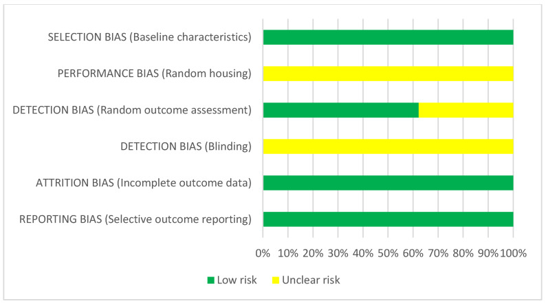
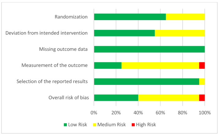
The regenerative endodontic procedure (REP) represents a treatment option for immature necrotic teeth with a periapical lesion. Currently, this therapy has a wide field of pre-clinical and clinical applications, but no standardization exists regarding successful criteria. Thus, by analysis of animal and human studies, the aim of this systematic review was to highlight the main characteristics of the tissue generated by REP. A customized search of PubMed, EMBASE, Scopus, and Web of Science databases from January 2000 to January 2022 was conducted. Seventy-five human and forty-nine animal studies were selected. In humans, the evaluation criteria were clinical 2D and 3D radiographic examinations. Most of the studies identified a successful REP with an asymptomatic tooth, apical lesion healing, and increased root thickness and length. In animals, histological and radiological criteria were considered. Newly formed tissues in the canals were fibrous, cementum, or bone-like tissues along the dentine walls depending on the area of the root. REP assured tooth development and viability. However, further studies are needed to identify procedures to successfully reproduce the physiological structure and function of the dentin-pulp complex.

Keywords: animal model; dentin-pulp complex regeneration; pulp injury; pulp necrosis; regenerative endodontics.


Conflict of interest statement

The authors declare no conflict of interest.

Figures

Figure 1
Figure 1
(a) Schema illustrating REP procedure. (A) Immature permanent incisor tooth exposed to trauma or caries. (B) Access cavity preparation and chemical debridement by using of irrigants and/or medicaments. (C) Bleeding induced by dental endodontic File to create a blood clot. (D) Restoration of the access cavity with a permanent restorative material covering the biomaterial in contact with blood clot or with collagen plug, PRF, PRP, etc. (E) Release of signaling molecules from biomaterial (growth factors, calcium silicate, depending on the material). They influence SCAPs (from apical papilla) and PDLs, including chemotaxis/cell migration, angiogenesis, neurogenesis, and differentiation into pulp/dentin complex. DPSCs (Dental Pulp Stem Cells); SCAPs (Stem Cells from Apical Papilla); PDLs (Periodontal Ligament cells) (Document of URP2496). (b) Different type of achievement of tissue regeneration determined by continued root development, increased dentinal wall thickness by cementum-like deposition, and apical closure. (A) Immature permanent incisor tooth after REP technique with ”pulp-like” tissue with ligamentous, fibrous aspect and apical closure with mineralized tissue: Cement-like or bone-like tissue; (B) ”pulp-like” tissue with ligamentous aspect and apical closure with mineralized tissue: Cement-like or bone-like tissue. Cementum island in the intracanal tissue; (C) the expectation of the pulp-like tissue with an increased root thickness and length, a decreasing apex diameter. Document of URP2496.
Figure 2
Figure 2
Flowchart of the article selection process.
Figure 3
Figure 3
Risk of bias assessment of REP in animal studies according to the Systematic Review Centre for Laboratory Animal Experimentation (SYRCLE): Authors’ judgment about each risk of bias item (green = low, yellow = unclear) [16,17,18,19,20,21,22,23,24,25,26,27,28,29,30,31,32,33,34,37,38,39,40,41,42,43,44,45,46,47,48,49,50,51,52,53,54,55,56,57,58,59,61,62,63,64,65].
Figure 3
Figure 3
Risk of bias assessment of REP in animal studies according to the Systematic Review Centre for Laboratory Animal Experimentation (SYRCLE): Authors’ judgment about each risk of bias item (green = low, yellow = unclear) [16,17,18,19,20,21,22,23,24,25,26,27,28,29,30,31,32,33,34,37,38,39,40,41,42,43,44,45,46,47,48,49,50,51,52,53,54,55,56,57,58,59,61,62,63,64,65].
Figure 4
Figure 4
Risk of bias assessment of REP in animal studies according to the Systematic Review Centre for Laboratory Animal Experimentation (SYRCLE) (green = low risk, yellow = unclear risk).
Figure 5
Figure 5
Risk of bias assessment evaluated according to the R.O.B 2.0. Authors’ judgment about the following items: Randomization, deviation from intended intervention, missing outcome data, measurement of the outcome, selection of the reported results, and overall risk of bias (green = low, yellow = unclear, red = high). [6,7,69,70,84,85,86,87,88,89,90,114,115,123,126,128,129,135].
Figure 6
Figure 6
Risk of bias assessment evaluated according to the R.O.B 2.0 tool. Randomization, deviation from intended intervention, missing outcome data, measurement of the outcome, selection of the reported results, and overall risk of bias (green = low, yellow = moderate, red = high).
Figure 7
Figure 7
Risk of bias summary: Authors’ judgement about each risk of bias item for each included non-randomized study (Robins I tool). [60,116,121,123,124,132,135].
Figure 8
Figure 8
Risk of bias assessment evaluated according to the ROBINS I tool. Confounding, selection of participants, classification of intervention, deviation from intended intervention, missing data, selection of the reported results, overall risk of bias (green = low, yellow = moderate, red = high).

References

    1. Diogenes A., Ruparel N.B. Regenerative Endodontic Procedures: Clinical Outcomes. Dent. Clin. N. Am. 2017;61:111–125. doi: 10.1016/j.cden.2016.08.004. - DOI - PubMed
    1. Galler K.M., Krastl G., Simon S., Van Gorp G., Meschi N., Vahedi B., Lambrechts P. European Society of Endodontology position statement: Revitalization procedures. Int. Endod. J. 2016;49:717–723. doi: 10.1111/iej.12629. - DOI - PubMed
    1. American Association of Endodontists Endodontic Diagnosis. 2013. [(accessed on 28 July 2022)]. Available online: https://www.aae.org/specialty/newsletter/endodontic-diagnosis/
    1. Kim S.G., Malek M., Sigurdsson A., Lin L.M., Kahler B. Regenerative endodontics: A comprehensive review. Int. Endod. J. 2018;51:1367–1388. doi: 10.1111/iej.12954. - DOI - PubMed
    1. Duggal M., Tong H.J., Al-Ansary M., Twati W., Day P.F., Nazzal H. Interventions for the endodontic management of non-vital traumatised immature permanent anterior teeth in children and adolescents: A systematic review of the evidence and guidelines of the European Academy of Paediatric Dentistry. Eur. Arch. Paediatr. Dent. 2017;18:139–151. doi: 10.1007/s40368-017-0289-5. - DOI - PMC - PubMed

Publication types

LinkOut - more resources