

Comparison between Immunocytochemistry, FISH and NGS for ALK and ROS1 Rearrangement Detection in Cytological Samples
- PMID: 36142468
- PMCID: PMC9502752
- DOI: 10.3390/ijms231810556
Comparison between Immunocytochemistry, FISH and NGS for ALK and ROS1 Rearrangement Detection in Cytological Samples
Abstract
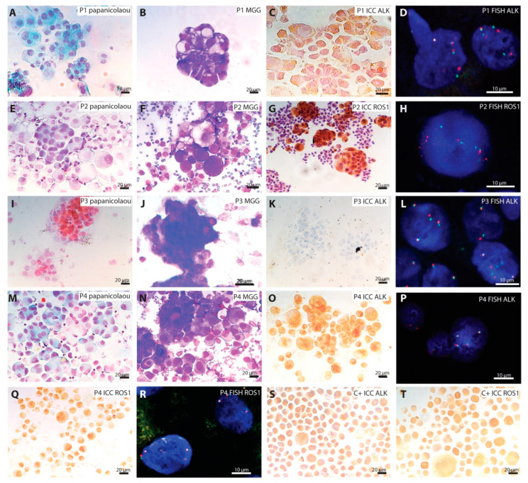
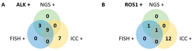
The detection of ROS1 and ALK rearrangements is performed for advanced-stage non-small cell lung cancer. Several techniques can be used on cytological samples, such as immunocytochemistry (ICC), fluorescence in situ hybridization (FISH) and, more recently, next-generation sequencing (NGS), which is gradually becoming the gold standard. We performed a retrospective study to compare ALK and ROS1 rearrangement results from immunocytochemistry, FISH and NGS methods from 131 cytological samples. Compared to NGS, the sensitivity and specificity of ICC were 0.79 and 0.91, respectively, for ALK, and 1 and 0.87 for ROS1. Regarding FISH, the sensitivity and specificity were both at 1 for ALK and ROS1 probes. False-positive cases obtained by ICC were systematically corrected by FISH. When using ICC and FISH techniques, results are very close to NGS. The false-positive cases obtained by ICC are corrected by FISH, and the true-positive cases are confirmed. NGS has the potential to improve the detection of ALK and ROS1 rearrangements in cytological samples; however, the cost of this technique is still much higher than the sequential use of ICC and FISH.
Keywords: ALK; FISH; NGS; ROS1; adenocarcinoma; cytology; immunocytochemistry; lung cancer.
Conflict of interest statement
The authors declare no conflict of interest.
Figures
Similar articles
-
Next-generation Sequencing for ALK and ROS1 Rearrangement Detection in Patients With Non-small-cell Lung Cancer: Implications of FISH-positive Patterns.Clin Lung Cancer. 2019 Jul;20(4):e421-e429. doi: 10.1016/j.cllc.2019.02.008. Epub 2019 Feb 26. Clin Lung Cancer. 2019. PMID: 30898567
-
Multiplex fluorescence in situ hybridisation to detect anaplastic lymphoma kinase and ROS proto-oncogene 1 receptor tyrosine kinase rearrangements in lung cancer cytological samples.J Clin Pathol. 2020 Feb;73(2):96-101. doi: 10.1136/jclinpath-2019-206152. Epub 2019 Sep 27. J Clin Pathol. 2020. PMID: 31562206
-
Detection of clinically actionable gene fusions by next-generation sequencing-based RNA sequencing of non-small cell lung cancer cytology specimens: A single-center experience with comparison to fluorescence in situ hybridization.Cancer Cytopathol. 2024 Jan;132(1):41-49. doi: 10.1002/cncy.22766. Epub 2023 Sep 25. Cancer Cytopathol. 2024. PMID: 37747438
-
ALK and ROS1 testing on lung cancer cytologic samples: Perspectives.Cancer Cytopathol. 2017 Nov;125(11):817-830. doi: 10.1002/cncy.21899. Epub 2017 Jul 25. Cancer Cytopathol. 2017. PMID: 28743163 Review.
-
Molecular diagnosis in non-small-cell lung cancer: expert opinion on ALK and ROS1 testing.J Clin Pathol. 2022 Mar;75(3):145-153. doi: 10.1136/jclinpath-2021-207490. Epub 2021 Apr 19. J Clin Pathol. 2022. PMID: 33875457 Free PMC article. Review.
Cited by
-
Comparative Efficacy of ALK Inhibitors for Treatment-Naïve ALK-Positive Advanced Non-Small Cell Lung Cancer with Central Nervous System Metastasis: A Network Meta-Analysis.Int J Mol Sci. 2023 Jan 23;24(3):2242. doi: 10.3390/ijms24032242. Int J Mol Sci. 2023. PMID: 36768562 Free PMC article. Review.
-
A narrative review of methods for the identification of ALK fusions in patients with non-small cell lung carcinoma.Transl Lung Cancer Res. 2023 Jul 31;12(7):1549-1562. doi: 10.21037/tlcr-22-855. Epub 2023 Jul 11. Transl Lung Cancer Res. 2023. PMID: 37577307 Free PMC article. Review.
-
Alectinib in combination with bevacizumab as first-line treatment in ALK-rearranged non-small cell lung cancer (ALEK-B): a single-arm, phase 2 trial.Nat Commun. 2025 May 16;16(1):4553. doi: 10.1038/s41467-025-59744-9. Nat Commun. 2025. PMID: 40379690 Free PMC article. Clinical Trial.
-
Cytological Samples: An Asset for the Diagnosis and Therapeutic Management of Patients with Lung Cancer.Cells. 2023 Feb 27;12(5):754. doi: 10.3390/cells12050754. Cells. 2023. PMID: 36899890 Free PMC article.
-
Progress in clinical diagnosis and treatment of colorectal cancer with rare genetic variants.Cancer Biol Med. 2024 Jun 15;21(6):473-83. doi: 10.20892/j.issn.2095-3941.2024.0026. Cancer Biol Med. 2024. PMID: 38940668 Free PMC article. Review.
References
-
- Cancer Today. [(accessed on 29 April 2022)]. Available online: http://gco.iarc.fr/today/home.
-
- Lindeman N.I., Cagle P.T., Aisner D.L., Arcila M.E., Beasley M.B., Bernicker E.H., Colasacco C., Dacic S., Hirsch F.R., Kerr K., et al. Updated Molecular Testing Guideline for the Selection of Lung Cancer Patients for Treatment with Targeted Tyrosine Kinase Inhibitors: Guideline From the College of American Pathologists, the International Association for the Study of Lung Cancer, and the Association for Molecular Pathology. Arch. Pathol. Lab. Med. 2018;142:321–346. doi: 10.5858/arpa.2017-0388-CP. - DOI - PubMed
-
- Chen Y.-F., Hsieh M.-S., Wu S.-G., Chang Y.-L., Shih J.-Y., Liu Y.-N., Tsai M.-F., Tsai T.-H., Yu C.-J., Yang J.C.-H., et al. Clinical and the Prognostic Characteristics of Lung Adenocarcinoma Patients with ROS1 Fusion in Comparison with Other Driver Mutations in East Asian Populations. J. Thorac. Oncol. 2014;9:1171–1179. doi: 10.1097/JTO.0000000000000232. - DOI - PubMed
MeSH terms
Substances
LinkOut - more resources
Full Text Sources
Medical
