Review
. 2022 Sep 12;23(18):10572.
doi: 10.3390/ijms231810572.

Glial Cell-Mediated Neuroinflammation in Alzheimer's Disease

Affiliations
Review

Glial Cell-Mediated Neuroinflammation in Alzheimer's Disease

Nour F Al-Ghraiybah et al. Int J Mol Sci. .

Abstract

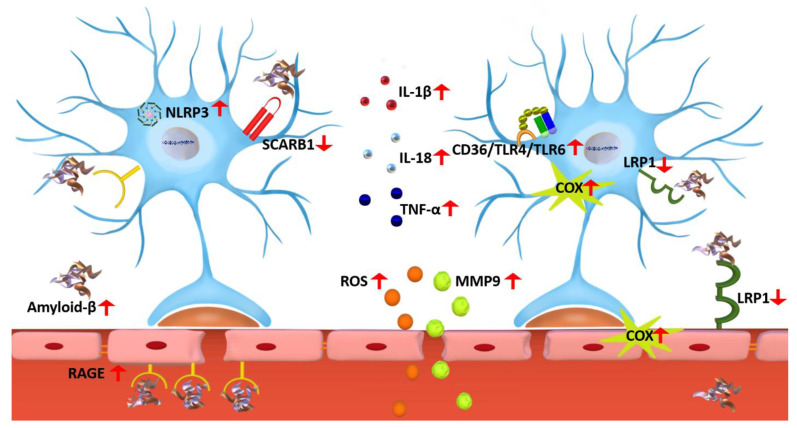
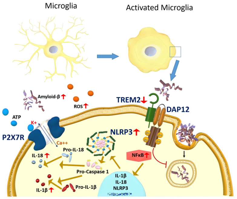
Alzheimer's disease (AD) is a progressive neurodegenerative disorder; it is the most common cause of dementia and has no treatment. It is characterized by two pathological hallmarks, the extracellular deposits of amyloid beta (Aβ) and the intraneuronal deposits of Neurofibrillary tangles (NFTs). Yet, those two hallmarks do not explain the full pathology seen with AD, suggesting the involvement of other mechanisms. Neuroinflammation could offer another explanation for the progression of the disease. This review provides an overview of recent advances on the role of the immune cells' microglia and astrocytes in neuroinflammation. In AD, microglia and astrocytes become reactive by several mechanisms leading to the release of proinflammatory cytokines that cause further neuronal damage. We then provide updates on neuroinflammation diagnostic markers and investigational therapeutics currently in clinical trials to target neuroinflammation.

Keywords: Alzheimer’s disease; astrocytes; clinical trials; diagnostic markers; microglia; neuroinflammation; therapeutics.


Conflict of interest statement

The authors declare no conflict of interest.

Figures

Figure 2
Figure 2
A representative scheme demonstrates astrocytes’ interaction with the BBB and the neuroinflammation effect mediated by astrocyte activation on the BBB function. In AD, reduced expression of astrocytic SCARB1 and LRP1 would decrease the clearance of Aβ. Reactive astrocytes produce a wide range of cytokines and other inflammatory markers such as ROS, RAGE, COX, and MMP9 due to NLRP3 inflammasomes and NF-κB pathways activation. In addition, TLR4 and TLR6 activation through binding to CD36 would increase proinflammatory cytokines secretion leading to neuroinflammation and BBB dysfunction. In AD, the expression of endothelium RAGE and LRP1 are upregulated and downregulated, respectively, which lead to reduced LRP1-mediated clearance of Aβ, and increased RAGE-mediated Aβ influx into the brain, which would further increase the vicious cycle of brain Aβ accumulation, astrocytes activation, and BBB dysfunction. Astrocytes, and microglia, could also be activated via TLRs. TLRs are pattern recognition receptors (PRR) that recognize and bind substrates and activate the immune response [186]. TLR could be expressed on the cell surface as TLR1, 2, 4, 5, 6, and 10 or intracellular as TLR 3, 7, 8, and 9, and their expression varies among immune cells [186]. In the immune cells of human AD brains, TLR mRNA is overexpressed except for TLR2, which is not changed [186]. Aβ is a substrate for TLR; upon binding, it stimulates its overexpression [187]. Recent research showed that while Aβ42 did not affect the expression of TLR3, it increased its reactivity in cultured microglial cells [188]. TLR4 antagonists could reduce neuroinflammation in AD, yet they might interfere with Aβ clearance by lowering the microglial phagocytotic ability of Aβ. In contrast, TLR4 agonists might have a beneficial role in Aβ clearance but not in reducing neuroinflammation [189]. Other researchers reported that the deletion of TLR2 in APPswe/PSEN1dE9 transgenic mice caused cognitive impairment, and increased anxiety, white matter damage, and brain atrophy in an Aβ-independent pathway [190], which collectively highlights the controversy regarding the role of TLR in AD [191].
Figure 1
Figure 1
Microglia activation in AD. Neuroinflammation in AD is associated with microglia activation that is mediated by increased Aβ, ROS, and ATP, which could activate P2X7R and downregulate TREM2. P2X7R activation would increase calcium influx, which leads to microglia activation and the release of inflammatory cytokines. In AD, reduced levels of TREM2 would impair Aβ phagocytosis by the microglia and thus increase Aβ brain parenchymal burden. Microglia activation is also associated with NF-κB and NLRP3 inflammasomes activation, which would activate caspase-1 and the secretion of proinflammatory cytokines IL-1β and IL-18.

Similar articles

Cited by

References

    1. Alzheimer’s Association 2018 Alzheimer’s disease facts, and figures. Alzheimer’s Dement. 2018;14:367–429. doi: 10.1016/j.jalz.2018.02.001. - DOI - PubMed
    1. Adav S.S., Sze S.K. Insight of brain degenerative protein modifications in the pathology of neurodegeneration and dementia by proteomic profiling. Mol. Brain. 2016;9:92. doi: 10.1186/s13041-016-0272-9. - DOI - PMC - PubMed
    1. Selkoe D.J. Toward a comprehensive theory for Alzheimer’s disease. Hypothesis: Alzheimer’s disease is caused by the cerebral accumulation and cytotoxicity of amyloid beta-protein. Ann. N. Y. Acad. Sci. 2000;924:17–25. doi: 10.1111/j.1749-6632.2000.tb05554.x. - DOI - PubMed
    1. Long J.M., Holtzman D.M. Alzheimer disease: An update on pathobiology and treatment strategies. Cell. 2019;179:312–339. doi: 10.1016/j.cell.2019.09.001. - DOI - PMC - PubMed
    1. Serrano-Pozo A., Frosch M.P., Masliah E., Hyman B.T. Neuropathological alterations in Alzheimer disease. Cold Spring Harb. Perspect. Med. 2011;1:a006189. doi: 10.1101/cshperspect.a006189. - DOI - PMC - PubMed