. 2022 Sep 13;23(18):10584.
doi: 10.3390/ijms231810584.

Recombinant Reg3α Prevents Islet β-Cell Apoptosis and Promotes β-Cell Regeneration

Affiliations

Recombinant Reg3α Prevents Islet β-Cell Apoptosis and Promotes β-Cell Regeneration

Luting Yu et al. Int J Mol Sci. .

Abstract

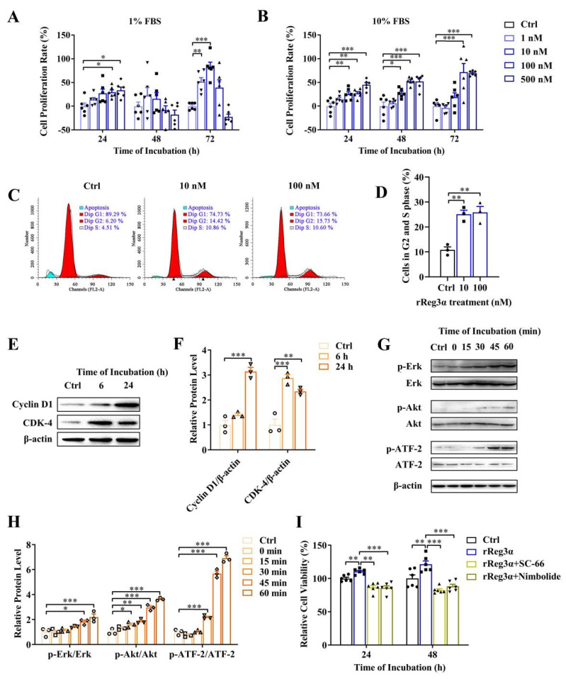
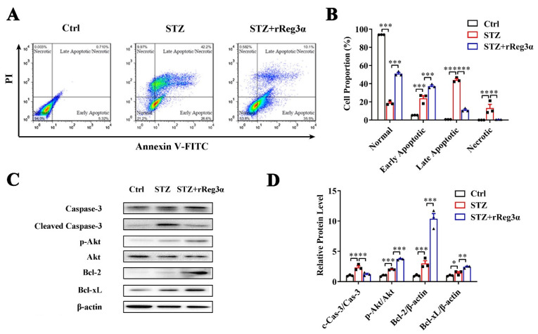
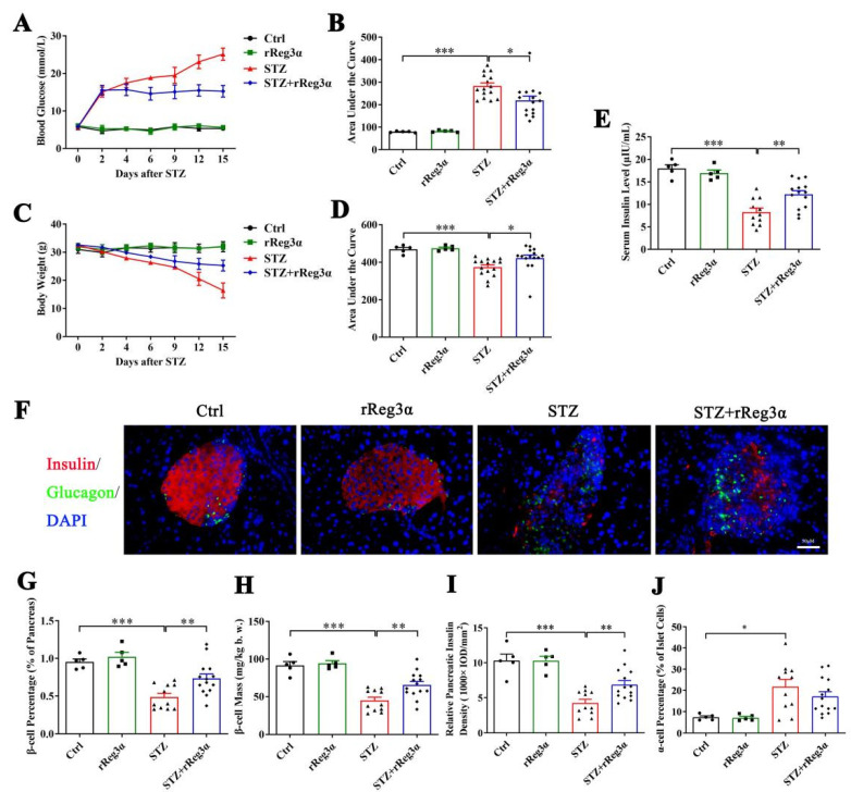
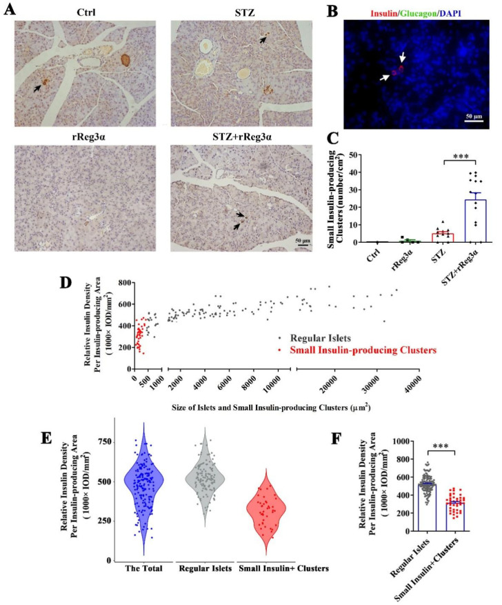
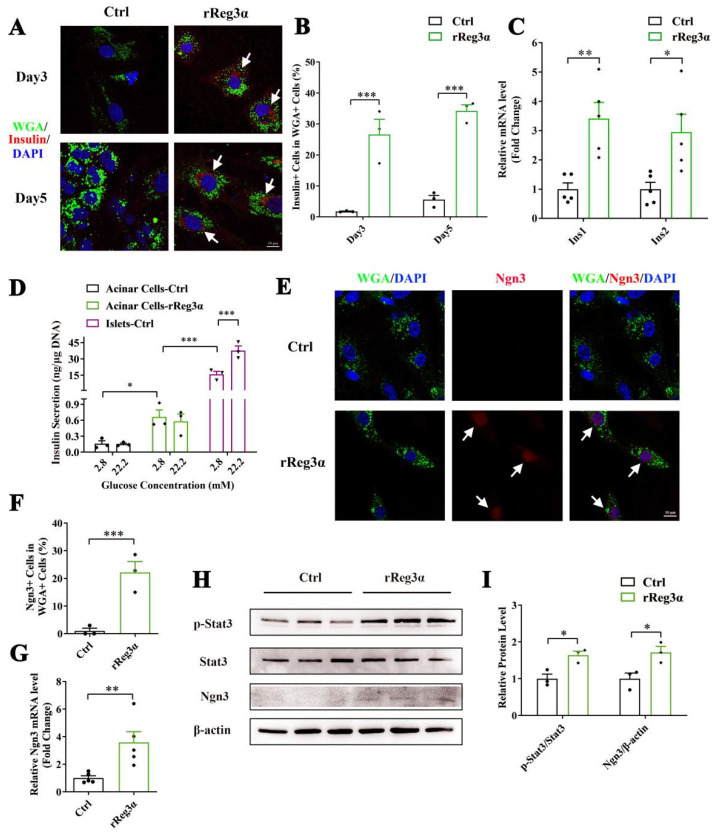
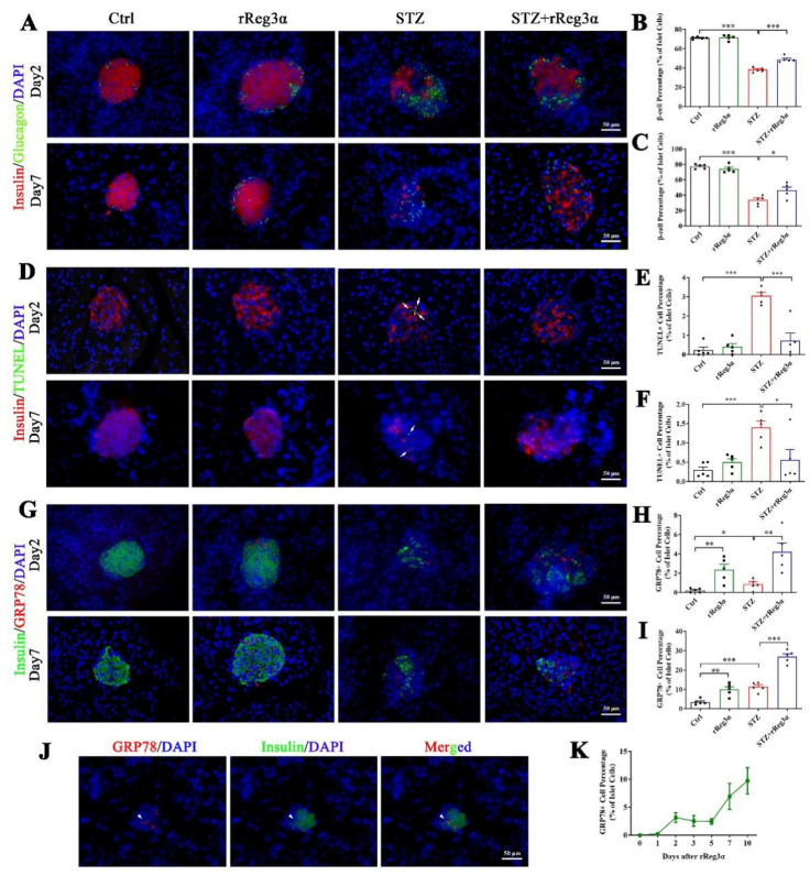
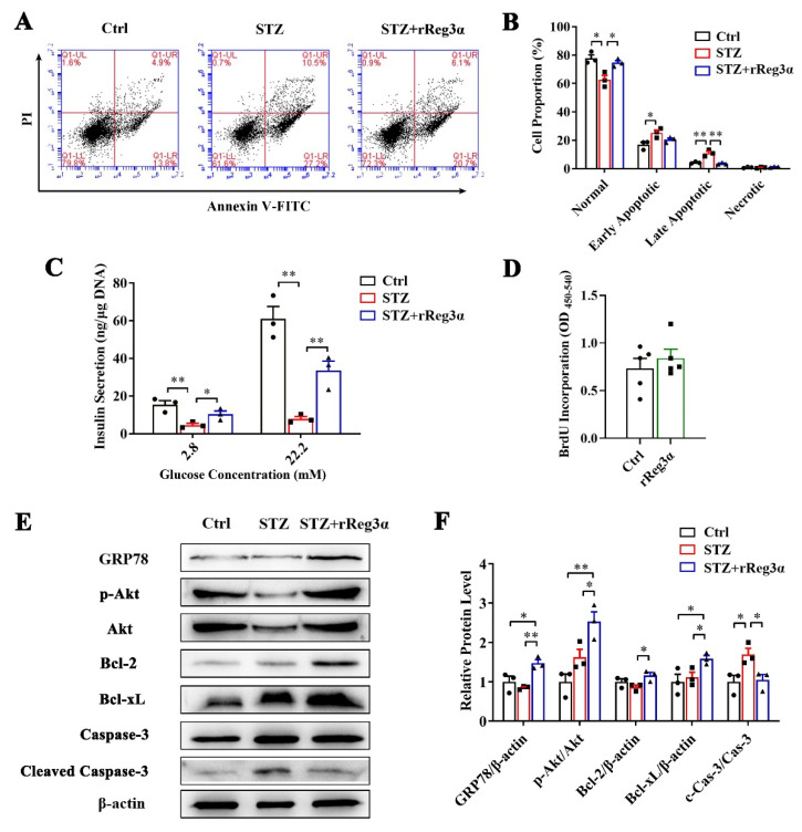
Progressive loss and dysfunction of islet β-cells has not yet been solved in the treatment of diabetes. Regenerating protein (Reg) has been identified as a trophic factor which is demonstrated to be associated with pancreatic tissue regeneration. We previously produced recombinant Reg3α protein (rReg3α) and proved that it protects against acute pancreatitis in mice. Whether rReg3α protects islet β-cells in diabetes has been elusive. In the present study, rReg3α stimulated MIN6 cell proliferation and resisted STZ-caused cell death. The protective effect of rReg3α was also found in mouse primary islets. In BALB/c mice, rReg3α administration largely alleviated STZ-induced diabetes by the preservation of β-cell mass. The protective mechanism could be attributed to Akt/Bcl-2/-xL activation and GRP78 upregulation. Scattered insulin-expressing cells and clusters with small size, low insulin density, and exocrine distribution were observed and considered to be neogenic. In isolated acinar cells with wheat germ agglutinin (WGA) labeling, rReg3α treatment generated insulin-producing cells through Stat3/Ngn3 signaling, but these cells were not fully functional in response to glucose stimulation. Our results demonstrated that rReg3α resists STZ-induced β-cell death and promotes β-cell regeneration. rReg3α could serve as a potential drug for β-cell maintenance in anti-diabetic treatment.

Keywords: GRP78; diabetes; regenerating protein; the islets; β-cell regeneration.


Conflict of interest statement

The authors declare no conflict of interest.

Figures

Figure 1
Figure 1
rReg3α stimulates MIN6 cell proliferation. (A) MTT test in MIN6 cells treated with a gradient concentration of rReg3α in 1% FBS. (B) MTT test in 10% FBS. * p < 0.05, ** p < 0.01, and *** p < 0.001 using one-way ANOVA, N = 6. (C) Cell cycle assay in MIN6 cells treated with rReg3α. Cells in S and G2 phases were considered proliferative. (D) Statistics of the proportion of proliferating cell in panel C. ** p < 0.01 using one-way ANOVA, N = 3. (E) Western blotting of CDK4 and cyclin D1 levels. (F) Densitometric quantification of the protein levels in panel E. The relative protein contents were corrected by β−actin. ** p < 0.01 and *** p < 0.001 using one-way ANOVA, N = 3. (G) Western blotting of Erk, Akt, and ATF−2 phosphorylation. (H) Densitometric quantification of the phosphorylated protein levels in panel G. The relative protein levels were corrected by the corresponding total proteins. * p < 0.05, ** p < 0.01, and *** p < 0.001 using one-way ANOVA, N = 3. (I) MTT test in MIN6 cells treated with rReg3α and inhibitors against Akt (SC−66) or Erk (Nimbolide). ** p < 0.01 and *** p < 0.001 using one-way ANOVA, N = 6.
Figure 2
Figure 2
rReg3α alleviates STZ-induced MIN6 cell apoptosis. (A) Apoptotic assessment in MIN6 cells treated with rReg3α prior to STZ. (B) Statistics of the cell proportion in panel A. ** p < 0.01 and *** p < 0.001 using one-way ANOVA, N = 3. (C) Western blotting of cleaved caspase-3, phosphorylated Akt, Bcl-2, and -xL levels in MIN6 cells. (D) Densitometric quantification of the protein levels in panel C. The relative cleaved caspase-3 and phosphorylated Akt contents were corrected by the corresponding total proteins and the relative Bcl-2 and -xL contents were corrected by β-actin. * p < 0.05, ** p < 0.01 and *** p < 0.001 using one-way ANOVA, N = 3.
Figure 3
Figure 3
rReg3α partially rescues STZ-induced diabetes in BALB/c mice. (A) Change in blood glucose level within 15 d after STZ. (B) AUC statistics of the blood glucose in panel A. (C) Change in weight loss. (D) AUC statistics of the bodyweight in panel C. * p < 0.05 and *** p < 0.001 using one-way ANOVA, N = 5, 5, 15, 15. (E) Change of serum insulin concentration 15 d after STZ. ** p < 0.01 and *** p < 0.001 using one-way ANOVA, N = 5, 5, 11, 14 (a few mice died before the endpoint). (F) Immunofluorescent staining to insulin and glucagon (400× magnification) 15 d after STZ. Cell nuclei were labeled with DAPI. A representative image is illustrated from each group. (G) Statistical analysis of β-cell percentage in the pancreas. (H) β-cell mass per bodyweight. (I) Semi-quantification of insulin content in the whole pancreas. The value of “relative pancreatic insulin density” indicated the total insulin density per total pancreatic area. (J) α-cell percentage in the islets. * p < 0.05, ** p < 0.01, and *** p < 0.001 using one-way ANOVA, N = 5, 5, 11, 14.
Figure 4
Figure 4
rReg3α induces insulin-producing cell neogenesis in the exocrine pancreas. (A) Micrographs of immunohistochemical staining to insulin (200× magnification) 15 d after STZ. A representative image is illustrated from each group. Up-left: the smallest islet, with a size of 425 μm2, observed in the pancreas from the control group. Insulin-producing cell clusters with the size less than 425 μm2 were considered to be neogenic. Black arrows indicate the putative neogenic insulin-producing cells. (B) Immunofluorescent staining to insulin and glucagon (400× magnification). A representative view is illustrated from all four groups. (C) Statistical analysis of the number of small insulin-producing clusters in panel A. *** p < 0.001 using one-way ANOVA, N = 5, 5, 11, 14. (D) Analysis of the regular islets (gray) and small insulin-producing clusters (red) in size and insulin density in the pancreas from STZ+rReg3α mice. (E) Violin plot of panel D. (F) Statistical analysis in panel E. *** p < 0.001 using one-way ANOVA. Regular Islets: insulin-stained structures with a size larger than 425 μm2, N = 133. Small Insulin+ Clusters: insulin-stained structures with a size less than 425 μm2, N = 42.
Figure 5
Figure 5
rReg3α promotes β-cell neogenesis in isolated acinar cells. (A) Confocal micrographs of immunofluorescent staining to insulin and WGA (630× magnification) in the isolated acinar cells treated with rReg3α at d 3 and 5. Cell nuclei were labeled with DAPI. A representative image is illustrated from each group. White arrows indicate the insulin+/WGA+ cells. (B) Statistical analysis of the insulin+/WGA+ cells in the total WGA+ cells in panel A. *** p < 0.001 using Student’s t-test, N = 3. (C) qRT-PCR analysis of Ins1 and Ins2 expression at d 3. * p < 0.05 and ** p < 0.01 using Student’s t-test, N = 5. (D) GSIS assay in the isolated acinar cells and primary islets at d 3. * p < 0.05 and *** p < 0.001 using one-way ANOVA, N = 3. (E) Confocal micrographs of immunofluorescent staining to Ngn3 and WGA (630× magnification) at d 3. Cell nuclei were labeled with DAPI. A representative image is illustrated from each group. White arrows indicate the Ngn3+/WGA+ cells. (F) Statistical analysis of the Ngn3+/WGA+ cells in the total WGA+ cells in panel E. *** p < 0.001 using Student’s t-test, N = 3. (G) qRT-PCR analysis of Ngn3 expression at d 3. ** p < 0.01 using Student’s t-test, N = 5. (H) Western blotting of Stat3 phosphorylation and Ngn3 level in the isolated acinar cells at d 3. (I) Densitometric quantification of the protein levels in panel H. The relative phosphorylated Stat3 content was corrected by the total Stat3 and the relative Ngn3 content was corrected by β-actin. * p < 0.05 using Student’s t-test, N = 3.
Figure 6
Figure 6
rReg3α upregulates GRP78 expression and prevents β-cell apoptosis. (A) Immunofluorescent staining to insulin and glucagon (400× magnification) at d 2 and 7 after STZ. Cell nuclei were labeled with DAPI. A representative image is illustrated from each group. (B,C) Statistical analysis of β-cell percentage in panel A. (D) Immunofluorescent staining to insulin and TUNEL (400× magnification) at d 2 and 7 after STZ. (E,F) Statistical analysis of TUNEL+ cell percentage in the islets in panel D. (G) Immunofluorescent staining to insulin and GRP78 (400× magnification) at d 2 and 7 after STZ. (H,I) Statistical analysis of GRP78high cell percentage in the islets in panel G. * p < 0.05, ** p < 0.01, and *** p < 0.001 using one-way ANOVA, N = 5. (J) Co-staining of insulin and GRP78high in a minority of endocrine cells. A representative image is illustrated from all groups. (K) Detection of GRP78high cells within 10 d after rReg3α treatment without STZ, N = 3.
Figure 7
Figure 7
rReg3α resists STZ-induced cell apoptosis in the isolated islets. (A) Apoptotic assessment in the isolated islets treated with rReg3α prior to STZ. (B) Statistics of the cell proportion in panel A. * p < 0.05, ** p < 0.01 using one-way ANOVA, N = 3. (C) GSIS assay, * p < 0.05 and ** p < 0.01 using one-way ANOVA, N = 3. (D) BrdU incorporation, N = 5. (E) Western blotting detection of GRP78, cleaved caspase-3, phosphorylated Akt, and Bcl-2, and -xL levels. (F) Densitometric quantification of the protein levels in panel E. The relative GRP78, Bcl-2, and -xL contents were corrected by β-actin and the relative phosphorylated Akt and cleaved caspase-3 contents were corrected by the corresponding total proteins. * p < 0.05 and ** p < 0.01 using one-way ANOVA, N = 3.

Similar articles

Cited by

References

    1. Retnakaran R., Rn A.E., Ye C., Harris S.B., Reichert S.M., McInnes N., Gerstein H.C., Thorpe K.E., Kramer C.K., Zinman B., et al. Short-term intensive insulin as induction and maintenance therapy for the preservation of beta-cell function in early type 2 diabetes (RESET-IT Main): A 2-year randomized controlled trial. Diabetes, Obes. Metab. 2021;23:1926–1935. doi: 10.1111/dom.14421. - DOI - PubMed
    1. Cheng C.W., Villani V., Buono R., Wei M., Kumar S., Yilmaz O.H., Cohen P., Sneddon J.B., Perin L., Longo V.D., et al. Fasting-Mimicking Diet Promotes Ngn3-Driven beta-Cell Regeneration to Reverse Diabetes. Cell. 2017;168:775–788.e12. doi: 10.1016/j.cell.2017.01.040. - DOI - PMC - PubMed
    1. Zhou Q., Melton D.A. Pancreas regeneration. Nature. 2018;557:351–358. - PMC - PubMed
    1. Robertson R.P. Islet transplantation a decade later and strategies for filling a half-full glass. Diabetes. 2010;59:1285–1291. doi: 10.2337/db09-1846. - DOI - PMC - PubMed
    1. Yonemura Y., Takashima T., Miwa K., Miyazaki I., Yamamoto H., Okamoto H. Amelioration of diabetes mellitus in partially depancreatized rats by poly(ADP-ribose) synthetase inhibitors. Evidence of islet B-cell regeneration. Diabetes. 1984;33:401–404. doi: 10.2337/diab.33.4.401. - DOI - PubMed