. 2022 Sep 13;23(18):10602.
doi: 10.3390/ijms231810602.

Effect of cx-DHED on Abnormal Glucose Transporter Expression Induced by AD Pathologies in the 5xFAD Mouse Model

Affiliations

Effect of cx-DHED on Abnormal Glucose Transporter Expression Induced by AD Pathologies in the 5xFAD Mouse Model

Jinho Kim et al. Int J Mol Sci. .

Abstract

Alzheimer's disease (AD) is a form of dementia associated with abnormal glucose metabolism resulting from amyloid-beta (Aβ) plaques and intracellular neurofibrillary tau protein tangles. In a previous study, we confirmed that carboxy-dehydroevodiamine∙HCl (cx-DHED), a derivative of DHED, was effective at improving cognitive impairment and reducing phosphorylated tau levels and synaptic loss in an AD mouse model. However, the specific mechanism of action of cx-DHED is unclear. In this study, we investigated how the cx-DHED attenuates AD pathologies in the 5xFAD mouse model, focusing particularly on abnormal glucose metabolism. We analyzed behavioral changes and AD pathologies in mice after intraperitoneal injection of cx-DHED for 2 months. As expected, cx-DHED reversed memory impairment and reduced Aβ plaques and astrocyte overexpression in the brains of 5xFAD mice. Interestingly, cx-DHED reversed the abnormal expression of glucose transporters in the brains of 5xFAD mice. In addition, otherwise low O-GlcNac levels increased, and the overactivity of phosphorylated GSK-3β decreased in the brains of cx-DHED-treated 5xFAD mice. Finally, the reduction in synaptic proteins was found to also improve by treatment with cx-DHED. Therefore, we specifically demonstrated the protective effects of cx-DHED against AD pathologies and suggest that cx-DHED may be a potential therapeutic drug for AD.

Keywords: Alzheimer’s disease; GSK-3β; O-GlcNac; cx-DHED; glucose transport.


Conflict of interest statement

The authors declare no conflict of interest. The funders had no role in the study design; collection, analyses, or interpretation of data; writing of the manuscript; or decision to publish the results.

Figures
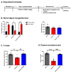
Figure 1
Figure 1
cx-DHED effects on cognitive impairment in 5xFAD mice. (A) The scheme showing the experimental procedure. After treatment with cx-DHED by intraperitoneal injection (1 mg/kg) for 2 months, behavioral testing was performed to determine the effect of cognitive behavior in 5xFAD mice. The behavior test was sequentially performed as follows: (B) novel object recognition test, (C) Y-maze test, and (D) passive avoidance test. Values are expressed as the mean ± SEM. ** p < 0.01, **** p < 0.0001 vs. WT-V, ### p < 0.001, #### p < 0.0001 vs. 5xFAD-V. Statistical analysis between the three groups was performed using one-way ANOVA, followed by Turkey’s post hoc test. Vehicle-treated wild-type (WT-V; n = 8), vehicle-treated 5xFAD (5xFAD-V; n = 6), and cx-DHED-treated 5xFAD (5xFAD-cx-DHED; n = 7) mice.
Figure 2
Figure 2
cx-DHED suppressed the formation of amyloid plaques. (A) Images showing the thioflavin-S stain localized to the amyloid plaques in the hippocampus (dentate gyrus) and cortex of 5xFAD mice. Quantification was determined as the number of plaques in the hippocampus (B) and cortex (C). Values are expressed as the mean ± standard error of the mean (5xFAD; n = 5, 5xFAD-cx-DHED; n = 5). Scale bars, 100 μm. * p < 0.05. Statistical analysis between the two groups was performed using the Student t-test.
Figure 3
Figure 3
cx-DHED reduced astrogliosis in the brain of 5xFAD mice. (A) Representative image of GFAP staining in the cortex to detect astrogliosis and (B) the quantification of GFAP immunofluorescence in the cortex of mice. (C) Western blot was performed to confirm the astrocyte expression level in the cortex of model mice. Values are expressed as the mean ± standard error of the mean (WT-V; n = 3, 5xFAD; n = 4, 5xFAD-cx-DHED; n = 4). β-actin was used as a loading control. **** p < 0.0001 vs. WT-V group; &&& p < 0.001, &&&& p < 0.0001 vs. 5xFAD-cxDHED group; # p < 0.05 and #### p < 0.0001 vs. 5xFAD-V group. Scale bars, 100 μm. Statistical analysis between the three groups was performed using the one-way analysis of variance, followed by Turkey’s post hoc test.
Figure 4
Figure 4
cx-DHED rescued the abnormal expressions of glucose transporters in the brain of 5xFAD mice. (A) Protein expression levels of glucose transporters (GLUT) in the cortex of mice. (B) GLUT1 (WT-V; n = 6, 5xFAD; n = 6, 5xFAD-cx-DHED; n = 6), (C) GLUT2 (WT-V; n = 6, 5xFAD; n = 6, 5xFAD-cx-DHED; n = 6), and (D) GLUT3 (WT-V; n = 5, 5xFAD; n = 5, 5xFAD-cx-DHED; n = 6) expression levels were normalized to β-actin. Values are expressed as the mean ± standard error of the mean. * p < 0.05, ** p < 0.01, and **** p < 0.0001 vs. WT-V group; # p < 0.05 and ### p < 0.001 vs. the 5xFAD-V group. Statistical analysis between the three groups was performed using the one-way analysis of variance, followed by Turkey’s post hoc test.
Figure 5
Figure 5
cx-DHED increased O-GlcNAcylation, but decreased phosphorylation of GSK-3β in the brain of 5xFAD mice. (A) Representative blot and quantification of O-GlcNac expression in the cortex of the mice (WT-V; n = 6, 5xFAD; n = 4, 5xFAD-cx-DHED; n = 4). (B) The representative blot and quantification of the phosphorylation of GSK-3β (pGSK-3β) and total GSK-3β (WT-V; n = 5, 5xFAD; n = 5, 5xFAD-cx-DHED; n = 4). β-actin was used as a loading control. Values are expressed as the mean ± SEM. ** p < 0.01 and **** p < 0.0001 vs. WT-V group; # p < 0.05 and #### p < 0.001 vs. 5xFAD-V group. Statistical analysis between the three groups was performed using the one-way analysis of variance, followed by Turkey’s post hoc test.
Figure 6
Figure 6
cx-DHED treatment alleviated loss of synaptic protein in the brain of 5xFAD mice. (A) The expression levels of PSD-95, a postsynaptic marker, and synaptophysin, presynaptic markers, were analyzed in the fraction of synaptic protein in the brain of mice. (B) Quantified values of PSD-95 and synaptophysin are expressed as the mean ± SEM (WT-V; n = 4, 5xFAD; n = 5, 5xFAD-cx-DHED; n = 5). β-actin was used as a loading control. ** p < 0.011 vs. WT-V group, # p < 0.05 vs. 5xFAD-V group. Statistical analysis between the three groups was performed using the one-way analysis of variance, followed by Turkey’s post hoc test.
Figure 7
Figure 7
Study summary. In the brain of 5xFAD mice, accumulation of amyloid plaques (Aβ) perturbs normal glucose metabolism by driving a reduction in glucose transporters 1 and 3 (GLUT1 and GLUT3, respectively) or overexpression of GLUT2 by upregulated reactive astrocytes. While phosphorylated GSK-3β was increased, O-GlcNac was diminished by dysfunctional glucose metabolism. Consequently, increased phosphorylated tau leads to synaptic loss and memory impairment. However, cx-DHED treatment attenuated the formation of amyloid plaques and upregulated O-GlcNac, as well as the level of GLUT in the brain of 5xFAD mice. Finally, cx-DHED treatment led to the reduction in phosphorylated tau and recovery of memory function.

Similar articles

Cited by

References

    1. Murphy M.P., LeVine H., 3rd Alzheimer’s disease and the amyloid-β peptide. J. Alzheimer’s Dis. 2010;19:311–323. doi: 10.3233/JAD-2010-1221. - DOI - PMC - PubMed
    1. Metaxas A., Kempf S.J. Neurofibrillary tangles in Alzheimer’s disease: Elucidation of the molecular mechanism by immunohistochemistry and tau protein phospho-proteomics. Neural Regen. Res. 2016;11:1579–1581. doi: 10.4103/1673-5374.193234. - DOI - PMC - PubMed
    1. Heneka M.T., Carson M.J., El Khoury J., Landreth G.E., Brosseron F., Feinstein D.L., Jacobs A.H., Wyss-Coray T., Vitorica J., Ransohoff R.M., et al. Neuroinflammation in Alzheimer’s disease. Lancet Neurol. 2015;14:388–405. doi: 10.1016/S1474-4422(15)70016-5. - DOI - PMC - PubMed
    1. Jackson J., Jambrina E., Li J., Marston H., Menzies F., Phillips K., Gilmour G. Targeting the Synapse in Alzheimer’s Disease. Front. Neurosci. 2019;13:735. doi: 10.3389/fnins.2019.00735. - DOI - PMC - PubMed
    1. Cummings J., Aisen P.S., DuBois B., Frolich L., Jack C.R., Jr., Jones R.W., Morris J.C., Raskin J., Dowsett S.A., Scheltens P. Drug development in Alzheimer’s disease: The path to 2025. Alzheimers Res. Ther. 2016;8:39. doi: 10.1186/s13195-016-0207-9. - DOI - PMC - PubMed