. 2022 Sep 13;23(18):10650.
doi: 10.3390/ijms231810650.

Cold Atmospheric Plasma Jet Treatment Improves Human Keratinocyte Migration and Wound Closure Capacity without Causing Cellular Oxidative Stress

Affiliations

Cold Atmospheric Plasma Jet Treatment Improves Human Keratinocyte Migration and Wound Closure Capacity without Causing Cellular Oxidative Stress

Aurélie Marches et al. Int J Mol Sci. .

Abstract

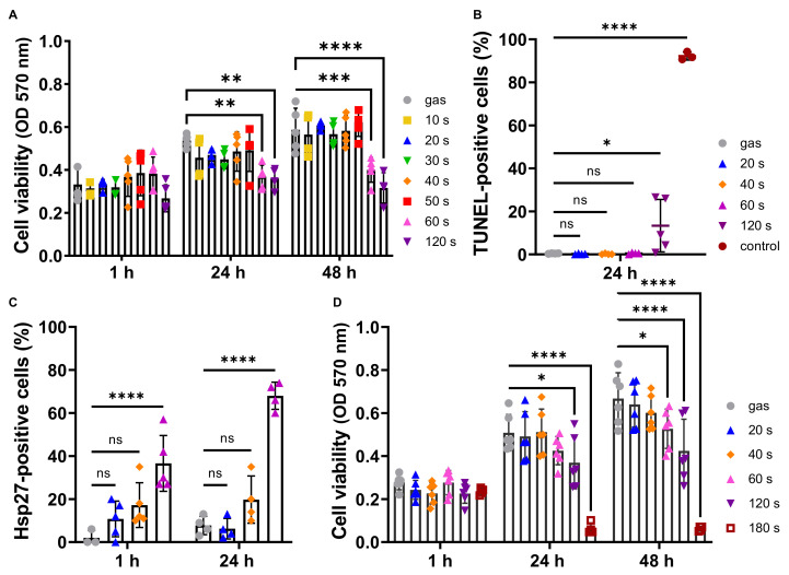
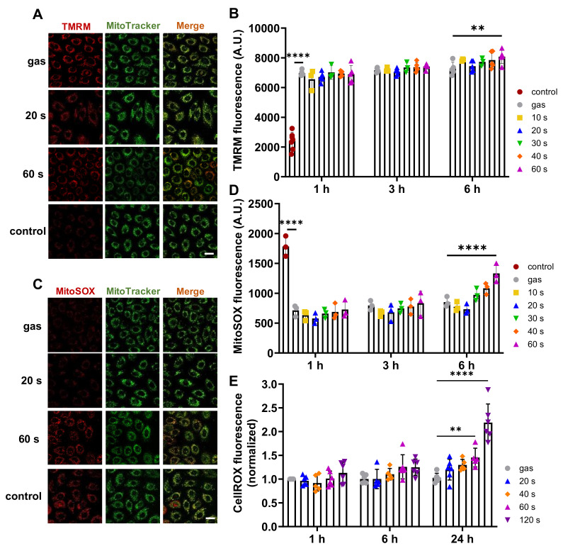
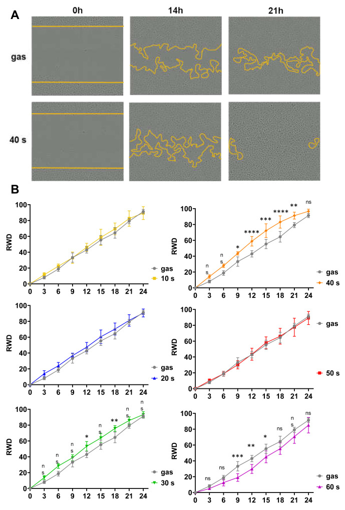
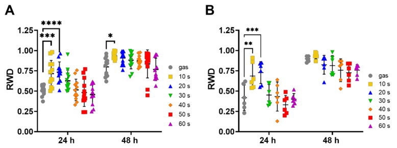
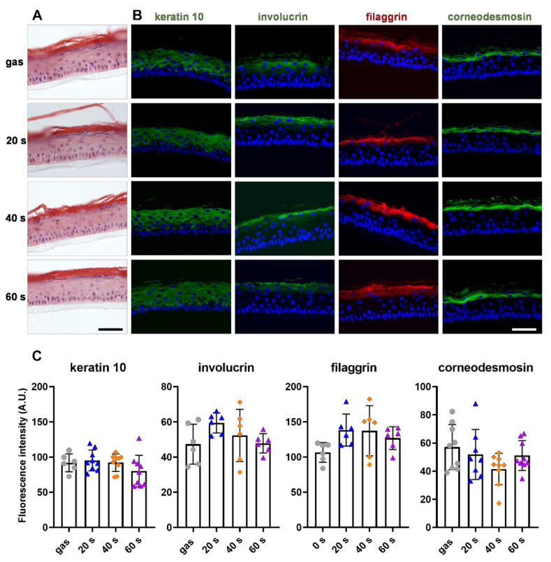
Cold Atmospheric Plasma (CAP) is an emerging technology with great potential for biomedical applications such as sterilizing equipment and antitumor strategies. CAP has also been shown to improve skin wound healing in vivo, but the biological mechanisms involved are not well known. Our study assessed a possible effect of a direct helium jet CAP treatment on keratinocytes, in both the immortalized N/TERT-1 human cell line and primary keratinocytes obtained from human skin samples. The cells were covered with 200 µL of phosphate buffered saline and exposed to the helium plasma jet for 10−120 s. In our experimental conditions, micromolar concentrations of hydrogen peroxide, nitrite and nitrate were produced. We showed that long-time CAP treatments (≥60 s) were cytotoxic, reduced keratinocyte migration, upregulated the expression of heat shock protein 27 (HSP27) and induced oxidative cell stress. In contrast, short-term CAP treatments (<60 s) were not cytotoxic, did not affect keratinocyte proliferation and differentiation, and did not induce any changes in mitochondria, but they did accelerate wound closure in vitro by improving keratinocyte migration. In conclusion, these results suggest that helium-based CAP treatments improve wound healing by stimulating keratinocyte migration. The study confirms that CAP could be a novel therapeutic method to treat recalcitrant wounds.

Keywords: cell migration; cold atmospheric plasma; keratinocytes; oxidative stress; reactive oxygen and nitrogen species; skin; wound healing.


Conflict of interest statement

The authors declare no conflict of interest. The funders had no role in the design of the study; in the collection, analyses, or interpretation of data; in the writing of the manuscript, or in the decision to publish the results.

Figures

Figure 1
Figure 1
CAP treatments of less than 40 s did not affect keratinocyte proliferation and viability. N/TERT-1 keratinocyte cell line (AC) and human primary keratinocytes (D) were exposed to CAP for the indicated periods of time. Cell viability was then determined using an MTT assay 1 h, 24 h and 48 h after CAP exposure (A,D), with cells treated with gas alone for 120 s as control; n ≥ 3. Induction of N/TERT-1 apoptosis was evaluated with a TUNEL assay 24 h after CAP treatments (B), 1 µM staurosporine being used as positive control; n = 3. Induction of heat stress was determined by immunocytochemistry on N/TERT-1 keratinocytes using an anti-HSP27 antibody, 1 h and 24 h after CAP exposure (C); n = 3. Error bars represent S.D.; *, p < 0.05; **, p < 0.005; ***, p < 0.001; ****, p < 0.0001; ns: not significant.
Figure 2
Figure 2
CAP treatment of less than 60 s did not affect N/TERT 1 mitochondria and intracellular ROS. N/TERT-1 cells were exposed to CAP for 10–60 s. Putative changes in mitochondrial membrane potential were determined by TMRM fluorescence assay 1 h, 3 h and 6 h post-treatment. Representative confocal microscopy images are shown (A) and fluorescence intensities were quantified by flow cytometry (B). We used 2 µM CCCP as positive control for membrane depolarization. n = 3. Mitochondrial dysfunctions were detected using the MitoSOX Red fluorescence assay. Representative confocal microscopy images are shown (C) and fluorescence intensities were quantified by flow cytometry (D). Two µM antimycin was used as positive control for mitochondrial anion superoxide production. n = 3. Changes in intracellular ROS levels were detected using green CellROX reagent by flow cytometry (E). Scale bars correspond to 20 µm. Error bars represent S.D.; **, p < 0.005; ****, p < 0.0001; ns: not significant.
Figure 3
Figure 3
Short-time CAP treatments of N/TERT-1 increased cell migration. Standardized scratches were made on a completely confluent cell monolayer, cells were exposed to CAP for between 10 s and 60 s, and images of the wound area were taken at hourly intervals for 24 h to quantify cell migration. Treatments with gas alone for 60 s was used as control. (A). Representative images of those taken immediately after the scratches (0 h), and 14 h and 21 h later. Yellow lines indicate the edges of the wound. (B). Quantitative analyses of scratch fillings were performed, and the results are expressed as relative wound density (RWD). The results corresponded to the measurements in 6 separate wells; n = 6, error bars represent S.D.; *, p < 0.05; **, p < 0.005; ***, p < 0.001; ****, p < 0.0001; ns: not significant.
Figure 4
Figure 4
Short term CAP treatments of human primary keratinocytes stimulated their migration. Scratches were made manually on a completely confluent monolayer of primary human keratinocytes pre-treated for 3 h (B) or not (A) with 10 µg/mL mitomycin, and cells were exposed to CAP for 10 to 60 s, as indicated. The plate wells were monitored by bright-field microscopy 24 h and 48 h after plasma treatments, and quantitative analyses of the scratch closing, expressed as relative wound density (RWD), were performed. Treatments with gas alone for 60 s were used as control. The results corresponded to experiments performed with keratinocytes from 3 different donors; n = 6, error bars represent S.D.; *, p < 0.05; **, p < 0.005; ***, p < 0.001; ****, p < 0.0001; ns: not significant.
Figure 5
Figure 5
CAP treatments did not affect primary keratinocyte differentiation. Human primary keratinocytes were exposed to the direct plasma jet for 20 s, 40 s and 60 s, and used to produce reconstructed epidermis at the air-liquid interface; treatment for 60 s with gas alone was used as control. The resulting epidermis were formalin-fixed and paraffin-embedded. (AC) Five µm sections were stained with H&E (A) and immunodetected with antibodies specific for keratin 10 (green), involucrin (green), filaggrin (red) and corneodesmosin (green) (B,C). Representative immunofluorescence images are shown in (B). Nucleus staining (DAPI) is in blue. Scale bars are 50 µm. Immunofluorescence signals were quantified using ImageJ (C). Error bars represent S.D.

References

    1. Sen C.K. Wound healing essentials: Let there be oxygen. Wound Repair Regen. 2009;17:1–18. doi: 10.1111/j.1524-475X.2008.00436.x. - DOI - PMC - PubMed
    1. Morton L.M., Phillips T.J. Wound healing and treating wounds: Differential diagnosis and evaluation of chronic wounds. J. Am. Acad. Dermatol. 2016;74:589–605. doi: 10.1016/j.jaad.2015.08.068. - DOI - PubMed
    1. Dunnill C., Patton T., Brennan J., Barrett J., Dryden M., Cooke J., Leaper D., Georgopoulos N.T. Reactive oxygen species (ROS) and wound healing: The functional role of ROS and emerging ROS-modulating technologies for augmentation of the healing process. Int. Wound J. 2017;14:89–96. doi: 10.1111/iwj.12557. - DOI - PMC - PubMed
    1. Pastar I., Stojadinovic O., Yin N.C., Ramirez H., Nusbaum A.G., Sawaya A., Patel S.B., Khalid L., Isseroff R.R., Tomic-Canic M. Epithelialization in Wound Healing: A Comprehensive Review. Adv. Wound Care. 2014;3:445–464. doi: 10.1089/wound.2013.0473. - DOI - PMC - PubMed
    1. Hiller J., Stratmann B., Timm J., Costea T.-C., Tschoepe D. Enhanced growth factor expression in chronic diabetic wounds treated by cold atmospheric plasma. Diabet. Med. 2022;39:e14787. doi: 10.1111/dme.14787. - DOI - PubMed