. 2022 Sep 16;23(18):10819.
doi: 10.3390/ijms231810819.

AST-487 Inhibits RET Kinase Driven TERT Expression in Bladder Cancer

Affiliations

AST-487 Inhibits RET Kinase Driven TERT Expression in Bladder Cancer

Neeraj Agarwal et al. Int J Mol Sci. .

Abstract

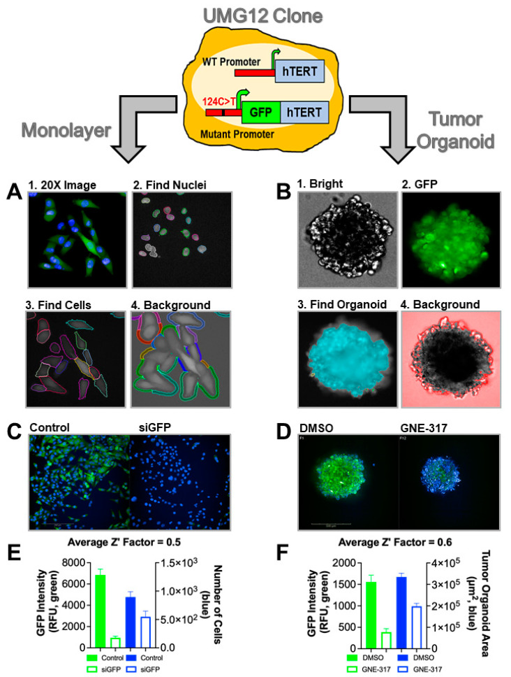
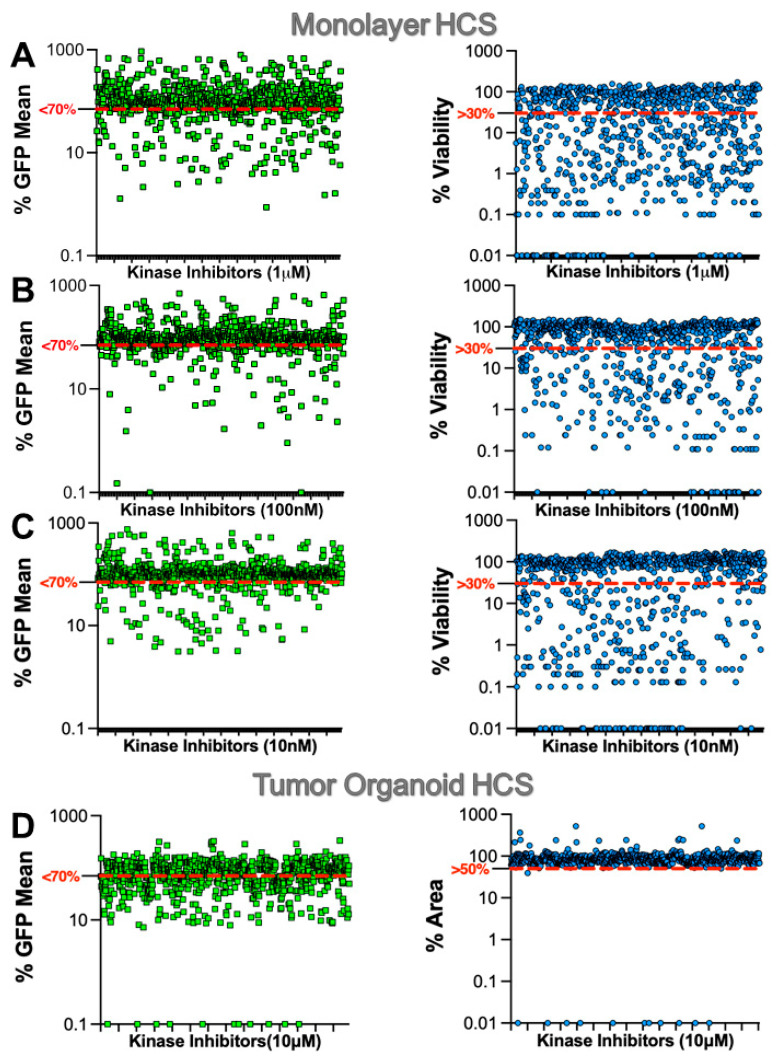
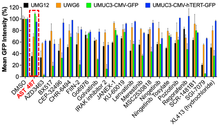
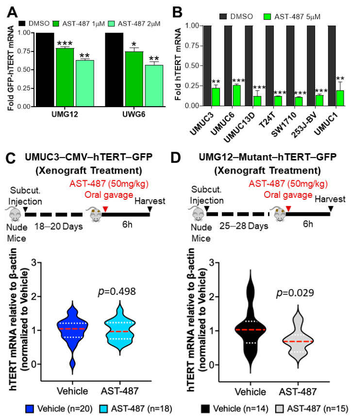
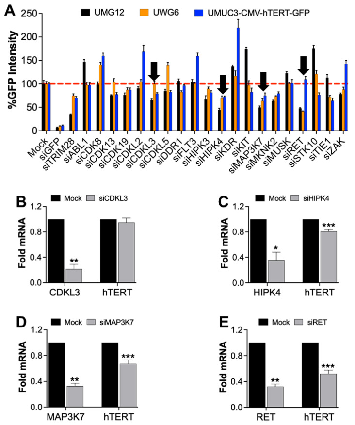
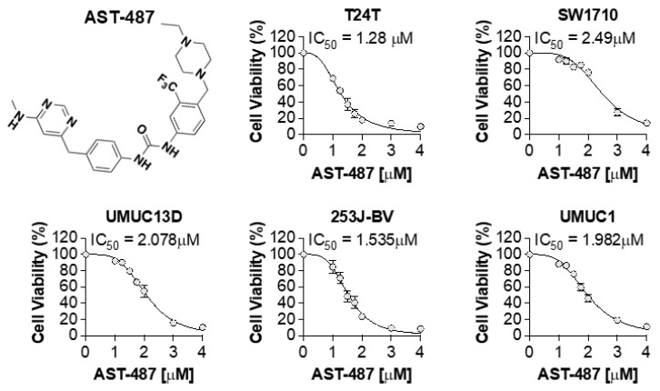
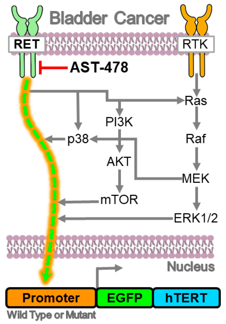
Mutations in the promoter of the human Telomerase Reverse Transcriptase (hTERT) gene are common and associated with its elevated expression in bladder cancer, melanoma, and glioblastoma. Though these mutations and TERT overexpression are associated with aggressive disease and poor outcome, an incomplete understanding of mutant TERT regulation limits treatment options directed at this gene. Herein, we unravel a signaling pathway that leads to upregulated hTERT expression resulting from the -124 bp promoter mutation, the most frequent variant across human cancer. We employed engineered bladder cancer cells that harbor a GFP insertion at the TSS region on -124 hTERT promoter for high-content screening drug discovery using a focused library of ~800 kinase inhibitors. Studies using in vitro and in vivo models prioritized AST-487, an inhibitor of the wild-type, and mutant RET (rearranged during transfection) proto-oncogene as a novel drug inhibitor of both wild-type and mutant promoter-driven hTERT expression. We also identified the RET kinase pathway, targeted by AST-487, as a novel regulator of mutant hTERT promoter-driven transcription in bladder cancer cells. Collectively, our work provides new potential precision medicine approaches for cancer patients with upregulated hTERT expression, perhaps, especially those harboring mutations in both the RET gene and the hTERT promoter, such as in thyroid cancer.

Keywords: RET; TERT; telomerase.


Conflict of interest statement

The authors declare no conflict of interest.

Figures

Figure 1
Figure 1
HCS assay validation in UMG12 cell models. UMUC3 BLCA cells engineered with a mutant promoter-driven GFP-hTERT fusion reporter cell line (UMG12) were developed and validated for HCS drug discovery in cells cultured as monolayers or tumor organoids. (A) monolayer and (B) tumor organoid Step-wise high-content analysis workflow of images acquired after HCS. (C) Monolayer and (D) Tumor organoid representative images of HCS controls showing GFP intensity after the PI3K inhibitor GNE-317 (1 μM, +control), a known inhibitor of hTERT expression. (E) Monolayer and (F) Tumor organoid Z’-factor analysis demonstrating valid controls for HCS.
Figure 2
Figure 2
HCS of kinase inhibitors in UMG12 cell models. UMG12s were used to screen a focused library of kinase inhibitors in cells cultured as (AC) monolayers or (D) tumor organoids. HCS of monolayer cells were treated with 3 different concentrations of kinase inhibitor library as indicated. The hit limit for mean GFP intensity was set to less than 70% (red dashed line) for each HCS condition. In addition, cell viability was multiplexed with GFP mean intensity with a hit limit set at great than 30% (monolayer) and 50% (tumor organoid) viability, respectively. Both GFP and viability hit limits were used to select hits from HCS.
Figure 3
Figure 3
HCS identified 21 hits targeting 16 kinases with no known association to hTERT. The Mean GFP intensity was measured in UMG12, UWG6, UMUC3-CMV-GFP, and UMUC3-CMV-hTERT-GFP cells treated with 21 kinase inhibitors. Red dotted line box indicate the effect of AST-487 on cell proliferation.
Figure 4
Figure 4
Validation of AST-487 for effect on hTERT expression in vitro and in vivo. (A) GFP-hTERT mRNA expression from UMG12 and UWG6 cells were treated with AST-487, as indicated, for 16 h, followed by qRT-PCR analysis. (B) hTERT mRNA expression from different BLCA cells was treated with AST-487 for 16 h, followed by qRT-PCR analysis. Schematics showing (C) UMUC3-CMV-hTERT-GFP and (D) UMG12 tumor xenografts development and treatment with AST-487 in nude mice as indicated on the top of the graph panels. Tumors were harvested, followed by qRT-PCR analysis. hTERT mRNA expression is shown (bottom of the graph panels) from (C) UMUC3-CMV-hTERT-GFP and (D) UMG12 xenografts 6 h post AST-487 treatment. The significance is indicated as: * p < 0.05, ** p < 0.01, *** p < 0.001.
Figure 5
Figure 5
Kinase target validation for AST-487 affecting hTERT expression. (A) AST-487 inhibits several kinases, which were transiently knocked down in cells as indicated and monitored for GFP intensity after 72 h. Kinases affecting hTERT transcription are shown with black arrows. (BE) T24T BLCA cells were treated with siRNAs as indicated, and the knockdown of genes and effect on hTERT mRNA was confirmed by qRT-PCR. The student’s t-test indicated significance: * p < 0.05, ** p < 0.01, *** p < 0.001.
Figure 6
Figure 6
AST-487 cell viability dose-response studies. The chemical structure of AST-487. BLCA cell lines were treated with various concentrations of AST-487 for 72 h measuring cell viability and IC50 values.
Figure 7
Figure 7
Summary of mechanisms regulating hTERT expression in BLCA. The results herein link the receptor tyrosine kinase RET as a key factor regulating hTERT expression in BLCA (green & orange dashed arrow). Parallel kinase signaling pathways known to regulate hTERT expression that are also activated by RET are shown with grey arrows and likely converge on upregulating hTERT expression. Upstream inhibition of RET attenuates hTERT promoter activation, including the more tumorigenic mutant hTERT promoter, regardless of the signaling pathways shown in grey.

Similar articles

References

    1. Dratwa M., Wysoczanska B., Lacina P., Kubik T., Bogunia-Kubik K. TERT-Regulation and Roles in Cancer Formation. Front. Immunol. 2020;11:589929. doi: 10.3389/fimmu.2020.589929. - DOI - PMC - PubMed
    1. Colebatch A.J., Dobrovic A., Cooper W.A. TERT gene: Its function and dysregulation in cancer. J. Clin. Pathol. 2019;72:281–284. doi: 10.1136/jclinpath-2018-205653. - DOI - PubMed
    1. Pestana A., Vinagre J., Sobrinho-Simoes M., Soares P. TERT biology and function in cancer: Beyond immortalisation. J. Mol. Endocrinol. 2017;58:R129–R146. doi: 10.1530/JME-16-0195. - DOI - PubMed
    1. Shay J.W., Bacchetti S. A survey of telomerase activity in human cancer. Eur. J. Cancer. 1997;33:787–791. doi: 10.1016/S0959-8049(97)00062-2. - DOI - PubMed
    1. Chiba K., Johnson J.Z., Vogan J.M., Wagner T., Boyle J.M., Hockemeyer D. Cancer-associated TERT promoter mutations abrogate telomerase silencing. eLife. 2015;4:e07918. doi: 10.7554/eLife.07918. - DOI - PMC - PubMed

MeSH terms