. 2022 Sep 16;23(18):10832.
doi: 10.3390/ijms231810832.

A Single Aspergillus fumigatus Gene Enables Ergothioneine Biosynthesis and Secretion by Saccharomyces cerevisiae

Affiliations

A Single Aspergillus fumigatus Gene Enables Ergothioneine Biosynthesis and Secretion by Saccharomyces cerevisiae

Sean Doyle et al. Int J Mol Sci. .

Abstract

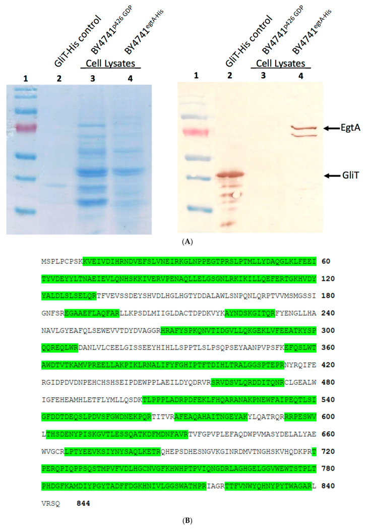
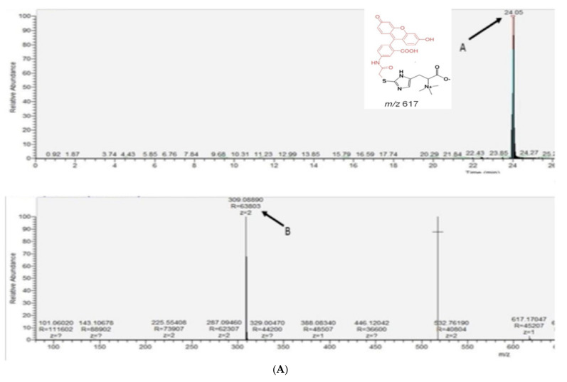
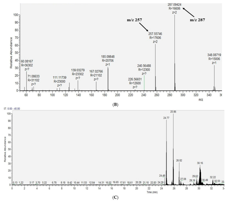
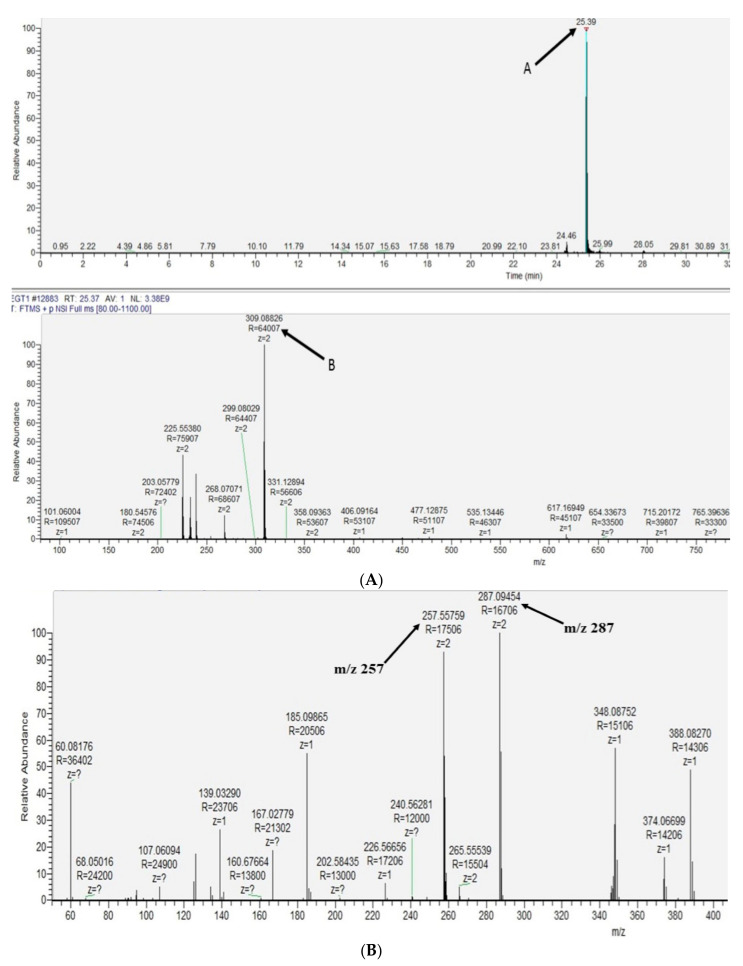
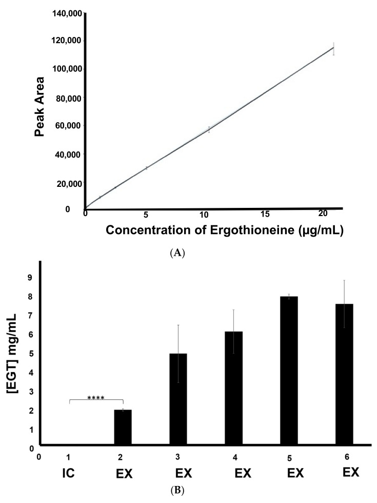
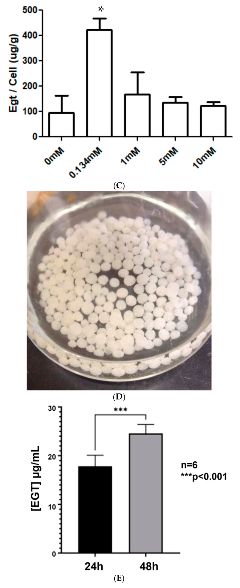
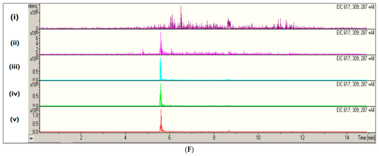
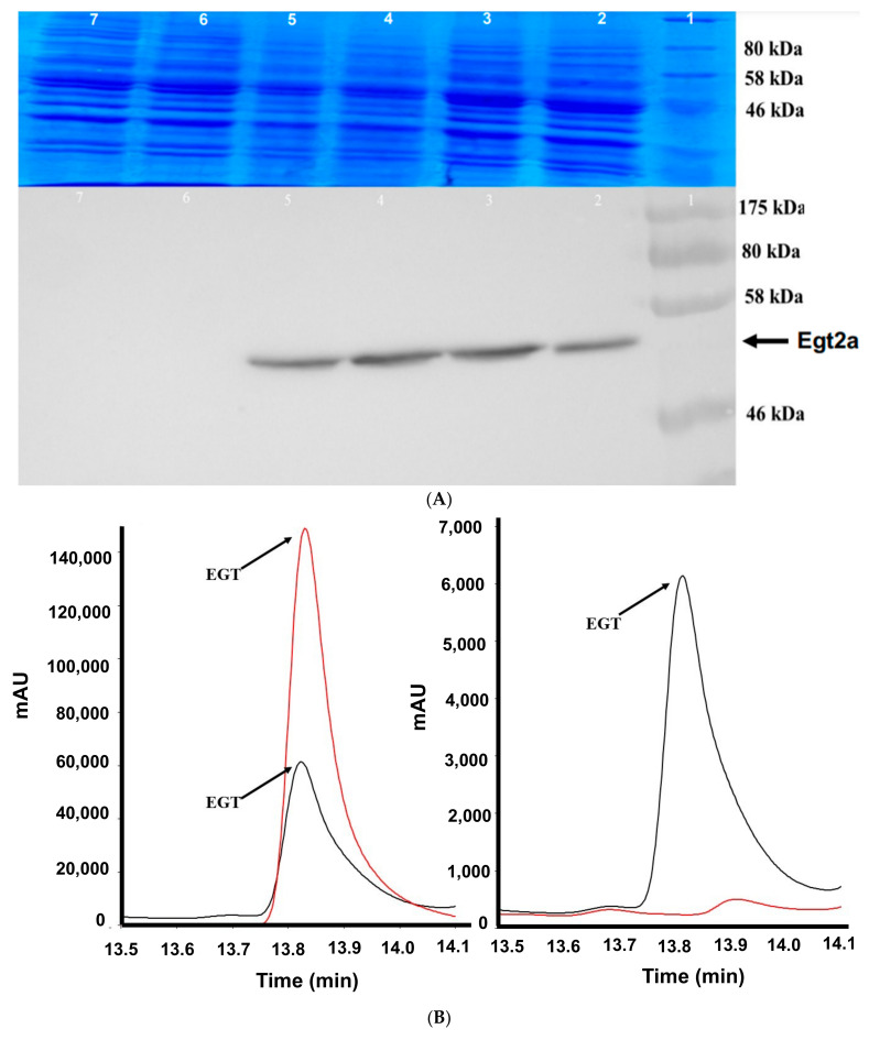
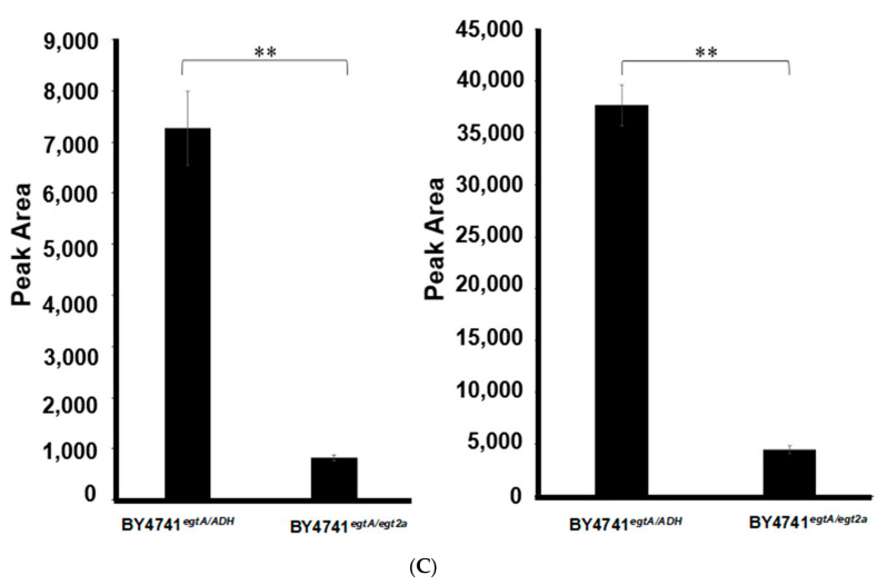
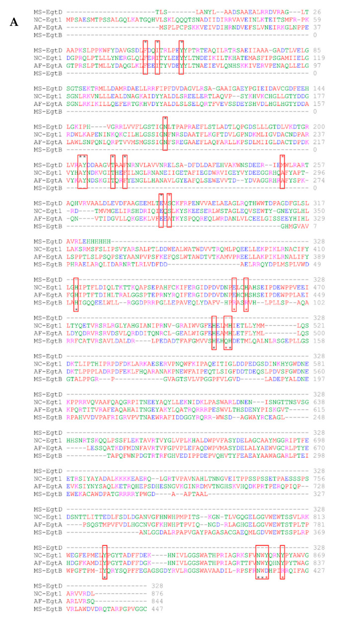
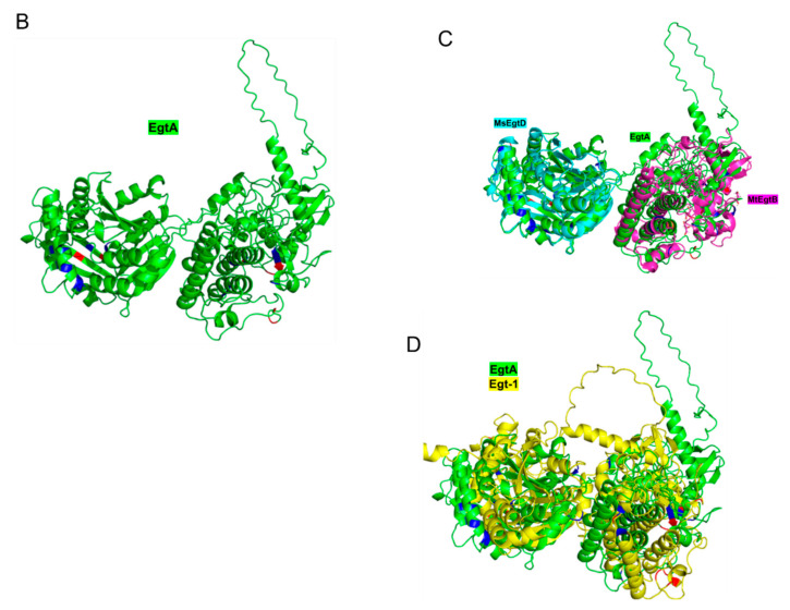
The naturally occurring sulphur-containing histidine derivative, ergothioneine (EGT), exhibits potent antioxidant properties and has been proposed to confer human health benefits. Although it is only produced by select fungi and prokaryotes, likely to protect against environmental stress, the GRAS organism Saccharomyces cerevisiae does not produce EGT naturally. Herein, it is demonstrated that the recombinant expression of a single gene, Aspergillus fumigatus egtA, in S. cerevisiae results in EgtA protein presence which unexpectedly confers complete EGT biosynthetic capacity. Both High Performance Liquid Chromatography (HPLC) and LC−mass spectrometry (MS) analysis were deployed to detect and confirm EGT production in S. cerevisiae. The localisation and quantification of the resultant EGT revealed a significantly (p < 0.0001) larger quantity of EGT was extracellularly present in culture supernatants than intracellularly accumulated in 96 h yeast cultures. Methionine addition to cultures improved EGT production. The additional expression of two candidate cysteine desulfurases from A. fumigatus was thought to be required to complete EGT biosynthesis, namely AFUA_2G13295 and AFUA_3G14240, termed egt2a and egt2b in this study. However, the co-expression of egtA and egt2a in S. cerevisiae resulted in a significant decrease in the observed EGT levels (p < 0.05). The AlphaFold prediction of A. fumigatus EgtA 3-Dimensional structure illuminates the bidomain structure of the enzyme and the opposing locations of both active sites. Overall, we clearly show that recombinant S. cerevisiae can biosynthesise and secrete EGT in an EgtA-dependent manner which presents a facile means of producing EGT for biotechnological and biomedical use.

Keywords: AlphaFold; ROS; antioxidant; cell factory; ergothioneine; redox stress.


Conflict of interest statement

The authors declare no conflict of interest.

Figures

Figure 1
Figure 1
(A) Western Blot confirming the expression of His-tagged EgtA in BY4741: Lane 1 = pre-stained protein ladder 10–170 kDa; Lane 2 = positive control recombinant GliT His-tag (36 kDa); Lane 3 = negative control BY4741p426 GDP-lysate; Lane 4 = BY4741egtA-His lysate. Western Blot visualised by 3,3′-diaminobenzidine; (B) EgtA identified from protein MS analysis of S. cerevisiae BY4741egtA-His cell lysate. EgtA was confirmed to be produced by BY474egt-His with 61% sequence coverage. Peptides from near the N- and C-termini are present and highlighted in green, indicating that EgtA was intact upon expression, although a smaller probably truncated protein can also be observed.
Figure 2
Figure 2
(A) Alkylated EGT in BY4741egtA-Hiscell lysate. LC–MS of BY4741egtA-His alkylated cell lysate revealed EGT presence at retention time of 24 min (A), including the double protonated form at m/z 309 (B); (B) daughter ions m/z 287 and m/z 257 of m/z 309 were identified also in cell lysate of BY4741-egtAwhich confirmed the intracellular presence of EGT; (C) neither m/z 309 or m/z 617 was present in the control culture BY4741p426-GPD, confirming EGT absence.
Figure 2
Figure 2
(A) Alkylated EGT in BY4741egtA-Hiscell lysate. LC–MS of BY4741egtA-His alkylated cell lysate revealed EGT presence at retention time of 24 min (A), including the double protonated form at m/z 309 (B); (B) daughter ions m/z 287 and m/z 257 of m/z 309 were identified also in cell lysate of BY4741-egtAwhich confirmed the intracellular presence of EGT; (C) neither m/z 309 or m/z 617 was present in the control culture BY4741p426-GPD, confirming EGT absence.
Figure 3
Figure 3
(A) The double protonated form of alkylated EGT (M + H)2+ = m/z 309 was identified in BY4741egtA culture supernatant at retention time of 25.39 min (A); (B) MS2 of m/z 309 yielded the expected daughter ions of m/z 309 (B), confirming extracellular alkylated EGT presence.
Figure 4
Figure 4
(A) Standard curve of labelled EGT. Standard curve generated using 9 EGT standards ranging from 0.08 to 20.83 µg/mL of EGT loaded onto column. Accuracy of standards was assessed by the R2 value = 0.9998 indicating a high degree of accuracy; (B) quantification of EGT production. Quantity of EGT in BY474-egtA grown in SC media: 1 = quantity of intracellular (IC) EGT (10.12 µg/L) after 24 h; 2 = quantity of extracellular (EX) EGT (1.91 mg/L) after 24 h, and at significantly **** (p ≤ 0.0001) higher amounts; 3 = quantity of extracellular EGT (4.89 mg/L) after 48 h; 4 = quantity of extracellular EGT (6.07 mg/L) after 72 h; 5 = quantity of extracellular EGT (7.93 mg/L) after 96 h; 6 = quantity of extracellular EGT (7.53 mg/L) after 120 h; (C) L-Met availability influences EGT production in recombinant S. cerevisiae. Amount of secreted EGT produced per weight of cells (μg/g) in BY4741egtA-His supernatants grown at a range of L-Met concentrations. Cultures grown at 0.134 mM L-Met show a significant * (p < 0.05) increase in EGT produced per gram of cells versus cultures grown at all other concentrations of L-Met; (D,E) EGT is released from immobilised recombinant S. cerevisiae–EgtA at 24 h and at significantly *** (p < 0.001) higher amounts at 48 h; (F) stacked EIC chromatograms following LC–MS analysis of (i) negative control, (ii) positive control (EGT (5 µg/mL), (iii) sample number 1 (iv) sample number 4, and (v) sample number 6. The peak visible at RT 5.6 min in samples (ii)–(v), inclusive, is EGT.
Figure 4
Figure 4
(A) Standard curve of labelled EGT. Standard curve generated using 9 EGT standards ranging from 0.08 to 20.83 µg/mL of EGT loaded onto column. Accuracy of standards was assessed by the R2 value = 0.9998 indicating a high degree of accuracy; (B) quantification of EGT production. Quantity of EGT in BY474-egtA grown in SC media: 1 = quantity of intracellular (IC) EGT (10.12 µg/L) after 24 h; 2 = quantity of extracellular (EX) EGT (1.91 mg/L) after 24 h, and at significantly **** (p ≤ 0.0001) higher amounts; 3 = quantity of extracellular EGT (4.89 mg/L) after 48 h; 4 = quantity of extracellular EGT (6.07 mg/L) after 72 h; 5 = quantity of extracellular EGT (7.93 mg/L) after 96 h; 6 = quantity of extracellular EGT (7.53 mg/L) after 120 h; (C) L-Met availability influences EGT production in recombinant S. cerevisiae. Amount of secreted EGT produced per weight of cells (μg/g) in BY4741egtA-His supernatants grown at a range of L-Met concentrations. Cultures grown at 0.134 mM L-Met show a significant * (p < 0.05) increase in EGT produced per gram of cells versus cultures grown at all other concentrations of L-Met; (D,E) EGT is released from immobilised recombinant S. cerevisiae–EgtA at 24 h and at significantly *** (p < 0.001) higher amounts at 48 h; (F) stacked EIC chromatograms following LC–MS analysis of (i) negative control, (ii) positive control (EGT (5 µg/mL), (iii) sample number 1 (iv) sample number 4, and (v) sample number 6. The peak visible at RT 5.6 min in samples (ii)–(v), inclusive, is EGT.
Figure 5
Figure 5
(A) Western Blot confirmation of Egt2a expression in BY4741egt2a-His: Lane 1 = protein ladder 175–177 kDa; Lanes 2–5 = independent samples of His-tagged Egt2a protein (50 kDa) from S. cerevisiae BY474egt2a-His; Lanes 6–7 = negative control BY4741egt2acell lysate; (B) EGT production in BY4741egtAegt2ais confirmed. Left panel shows EGT in BY4741egtAegt2a(retention time: 13.84 min (black) compared with the EGT standard 13.85 min (red)). Right panel shows EGT in BY4741egtAegt2a at a retention time of 13.84 min (black) compared with the BY4741p426-GPD p423 ADH where EGT is absent; (C) release of significantly less extracellular EGT from BY4741egtAegt2a than from BY4741egtAp423-ADH. Left panel shows culture supernatant EGT peak areas after 48 h and show significantly higher levels [**] (p = 0.0063) of EGT in BY4741egtAp423 ADHthan in BY4741egtAegt2a. Right panel reveals peak areas from EGT supernatant at 72 h which show significantly higher levels [**] (p = 0.0018) of EGT in BY4741egtAp423 ADHthan in BY4741egtAegt2a.
Figure 5
Figure 5
(A) Western Blot confirmation of Egt2a expression in BY4741egt2a-His: Lane 1 = protein ladder 175–177 kDa; Lanes 2–5 = independent samples of His-tagged Egt2a protein (50 kDa) from S. cerevisiae BY474egt2a-His; Lanes 6–7 = negative control BY4741egt2acell lysate; (B) EGT production in BY4741egtAegt2ais confirmed. Left panel shows EGT in BY4741egtAegt2a(retention time: 13.84 min (black) compared with the EGT standard 13.85 min (red)). Right panel shows EGT in BY4741egtAegt2a at a retention time of 13.84 min (black) compared with the BY4741p426-GPD p423 ADH where EGT is absent; (C) release of significantly less extracellular EGT from BY4741egtAegt2a than from BY4741egtAp423-ADH. Left panel shows culture supernatant EGT peak areas after 48 h and show significantly higher levels [**] (p = 0.0063) of EGT in BY4741egtAp423 ADHthan in BY4741egtAegt2a. Right panel reveals peak areas from EGT supernatant at 72 h which show significantly higher levels [**] (p = 0.0018) of EGT in BY4741egtAp423 ADHthan in BY4741egtAegt2a.
Figure 6
Figure 6
(A) Sequence alignment of bacterial and fungal ergothioneine biosynthetic enzymes, showing relevant active site amino acids (red boxes); (B) AlphaFold prediction of A. fumigatus EgtA 3D structure showing the bidomain enzyme arrangement, with active sites on opposing sites of the monomer (green); (C) MsEgtD methyltransferase (magenta) and MtEgtB sulphoxide synthase (pink), respectively, overlaid on A. fumigatus EgtA 3D structure; (D) the 3D structure of NcEgt-1 (yellow), predicted by AlphaFold, overlaid on the A. fumigatus EgtA 3D structure.
Figure 6
Figure 6
(A) Sequence alignment of bacterial and fungal ergothioneine biosynthetic enzymes, showing relevant active site amino acids (red boxes); (B) AlphaFold prediction of A. fumigatus EgtA 3D structure showing the bidomain enzyme arrangement, with active sites on opposing sites of the monomer (green); (C) MsEgtD methyltransferase (magenta) and MtEgtB sulphoxide synthase (pink), respectively, overlaid on A. fumigatus EgtA 3D structure; (D) the 3D structure of NcEgt-1 (yellow), predicted by AlphaFold, overlaid on the A. fumigatus EgtA 3D structure.

Similar articles

Cited by

References

    1. Seebeck F.P. In vitro reconstitution of Mycobacterial ergothioneine biosynthesis. J. Am. Chem. Soc. 2010;132:6632–6633. doi: 10.1021/ja101721e. - DOI - PubMed
    1. Sheridan K.J., Lechner B.E., Keeffe G.O., Keller M.A., Werner E.R., Lindner H., Jones G.W., Haas H., Doyle S. Ergothioneine Biosynthesis and Functionality in the Opportunistic Fungal Pathogen, Aspergillus fumigatus. Sci. Rep. 2016;6:35306. doi: 10.1038/srep35306. - DOI - PMC - PubMed
    1. Bello M.H., Barrera-Perez V., Morin D., Epstein L. The Neurospora crassa mutant NcDeltaEgt-1 identifies an ergothioneine biosynthetic gene and demonstrates that ergothioneine enhances conidial survival and protects against peroxide toxicity during conidial germination. Fungal Genet. Biol. 2012;49:160–172. doi: 10.1016/j.fgb.2011.12.007. - DOI - PubMed
    1. Cumming B.M., Chinta K.C., Reddy V.P., Steyn A.J.C. Role of Ergothioneine in Microbial Physiology and Pathogenesis. Antioxid Redox Signal. 2018;28:431–444. doi: 10.1089/ars.2017.7300. - DOI - PMC - PubMed
    1. Gamage A.M., Liao C., Cheah I.K., Chen Y., Lim D.R.X., Ku J.W.K., Chee R.S.L., Gengenbacher M., Seebeck F.P., Halliwell B., et al. The proteobacterial species Burkholderia pseudomallei produces ergothioneine, which enhances virulence in mammalian infection. FASEB J. 2018;32:6395–6409. doi: 10.1096/fj.201800716. - DOI - PubMed

LinkOut - more resources