. 2022 Sep 17;23(18):10860.
doi: 10.3390/ijms231810860.

Inhibition of FGFR Signaling by Targeting FGF/FGFR Extracellular Interactions: Towards the Comprehension of the Molecular Mechanism through NMR Approaches

Affiliations

Inhibition of FGFR Signaling by Targeting FGF/FGFR Extracellular Interactions: Towards the Comprehension of the Molecular Mechanism through NMR Approaches

Katiuscia Pagano et al. Int J Mol Sci. .

Abstract

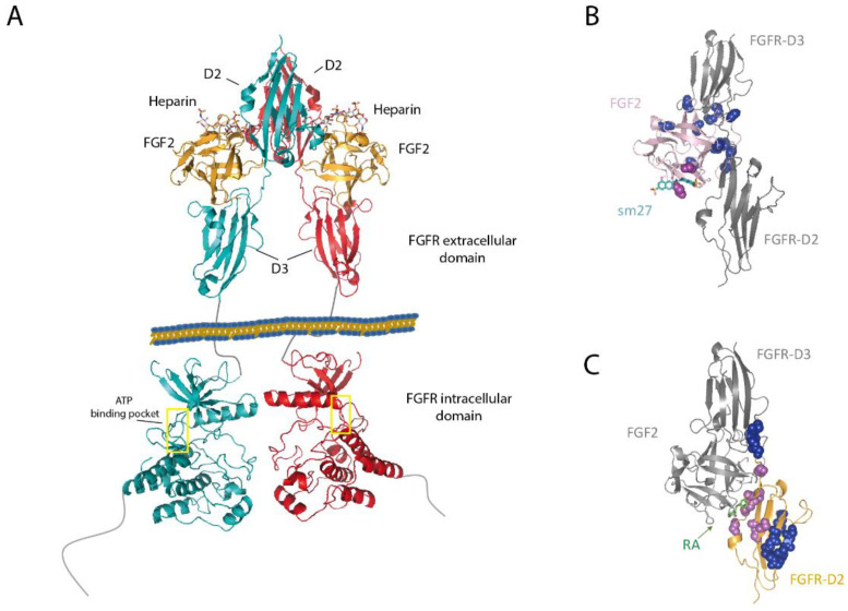
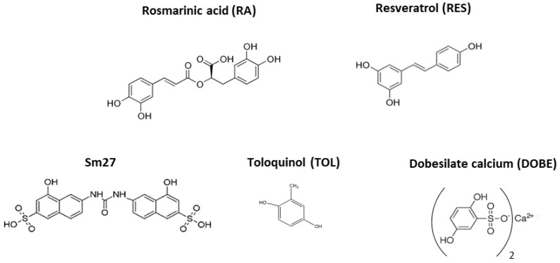
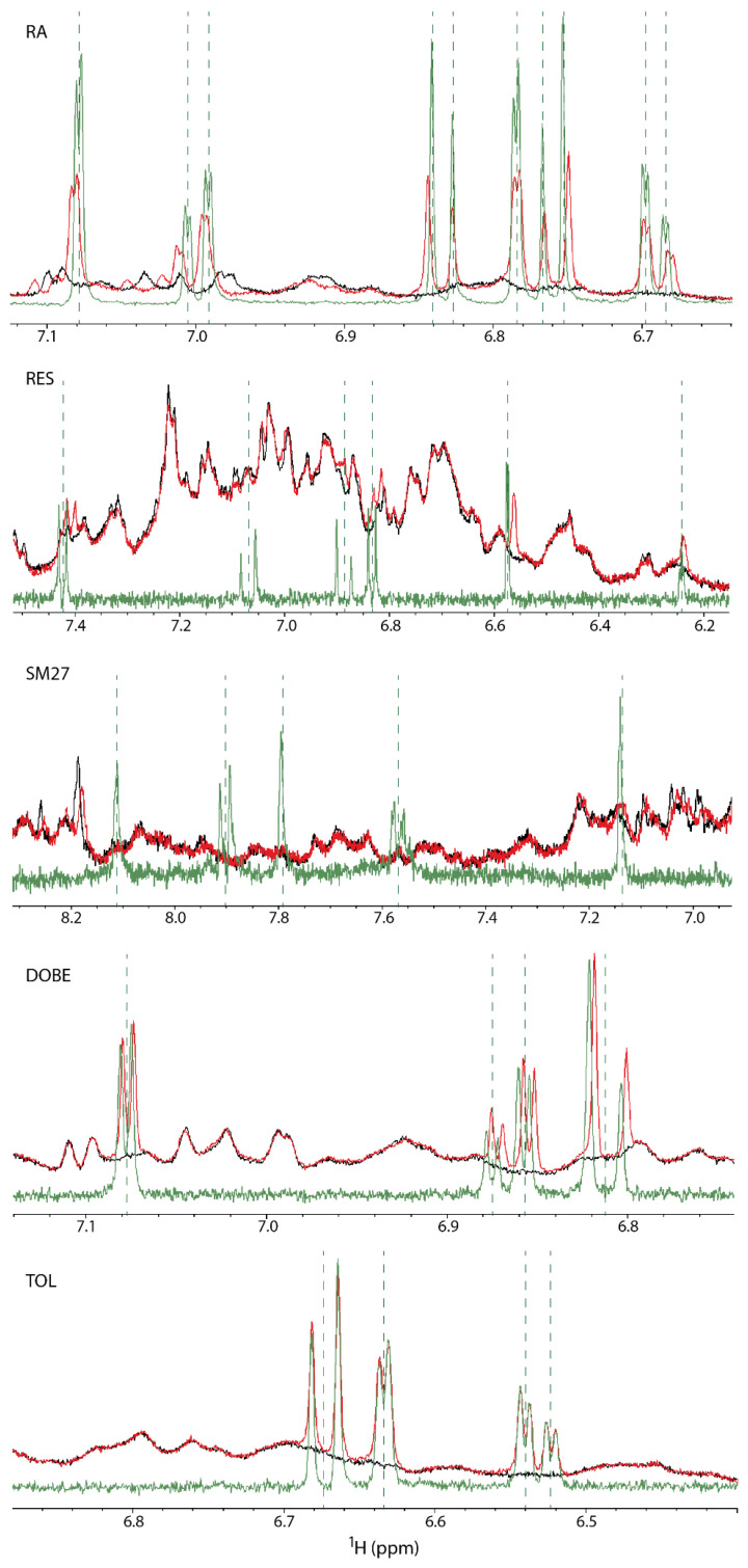
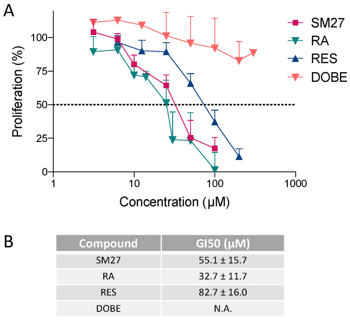
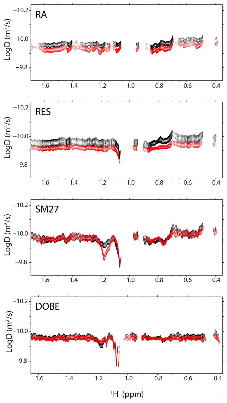
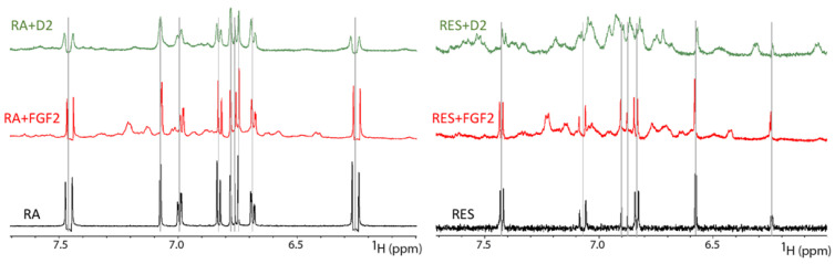
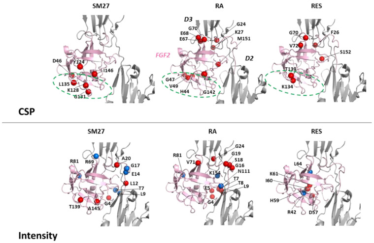
NMR-based approaches play a pivotal role in providing insight into molecular recognition mechanisms, affording the required atomic-level description and enabling the identification of promising inhibitors of protein-protein interactions. The aberrant activation of the fibroblast growth factor 2 (FGF2)/fibroblast growth factor receptor (FGFR) signaling pathway drives several pathologies, including cancer development, metastasis formation, resistance to therapy, angiogenesis-driven pathologies, vascular diseases, and viral infections. Most FGFR inhibitors targeting the intracellular ATP binding pocket of FGFR have adverse effects, such as limited specificity and relevant toxicity. A viable alternative is represented by targeting the FGF/FGFR extracellular interactions. We previously identified a few small-molecule inhibitors acting extracellularly, targeting FGFR or FGF. We have now built a small library of natural and synthetic molecules that potentially act as inhibitors of FGF2/FGFR interactions to improve our understanding of the molecular mechanisms of inhibitory activity. Here, we provide a comparative analysis of the interaction mode of small molecules with the FGF2/FGFR complex and the single protein domains. DOSY and residue-level NMR analysis afforded insights into the capability of the potential inhibitors to destabilize complex formation, highlighting different mechanisms of inhibition of FGF2-induced cell proliferation.

Keywords: DOSY; NMR; allosteric inhibitors; dobesilate; fibroblast growth factor; resveratrol; rosmarinic acid.


Conflict of interest statement

The authors declare no conflict of interest.

Figures

Figure 1
Figure 1
Structural features of the extracellular FGFR portion in complex with FGF2 and heparin, as well as the intracellular portion, including the ATP binding pocket (yellow rectangles), as derived from 1FQ9 [11] and 1FGI [8] PDB data, respectively (A). SM27 (B) and rosmarinic acid (RA) (C) binding sites (pink spheres) on FGF2 and FGFR-D2, respectively. Regions affected by long-range dynamic perturbations along the FGF2/FGFR interface region are highlighted with blue spheres.
Scheme 1
Scheme 1
The natural polyphenolic compound, rosmarinic acid (RA, Scheme 1 and Figure 1C), interacts with FGFR, inhibiting FGFR phosphorylation and FGF2-induced endothelial cell proliferation [26]. A variety of NMR approaches provide a molecular-level description of the interactions of RA with the FGF2/FGFR-D2 complex and both FGF2 and FGFR-D2 separately, showing that RA binds the FGFR-D2 domain and destabilizes the complex through direct and allosteric perturbations [24].
Figure 2
Figure 2
Comparison of 1D 1H spectra of small molecules, as indicated in the upper left, in the presence (red, FGF2:D2:small molecule 1:1:4) or absence (green) of the FGF2/D2 complex. The spectrum of the FGF2/D2 complex ([FGF2] = [D2] = 0.2 mM) is shown in black as a reference. Vertical dashed lines are positioned at the frequencies of the free-ligand resonances.
Figure 3
Figure 3
Effect of RA, RES, SM27, and DOBE on bovine aortic endothelial cells (BAEC). (A) Concentration-dependent inhibition of FGF2-induced BAEC proliferation by the compounds tested at the indicated concentration. Data are expressed as the percentage of control proliferation (mean and SEM). (B) GI50 values (in µM) for inhibition of FGF2-induced BAEC proliferation. N.A., not applicable; GI50 was not reached for DOBE.
Figure 4
Figure 4
Overlay of 2D NMR DOSY spectra of the FGF2/D2 complex ([FGF2] = [D2] = 0.2 mM) (black) and FGF2/D2 in the presence of the library molecules (red, FGF2:D2:small molecule 1:1:4). In each panel, the DOSY recorded on the freshly prepared FGF2/D2 twin samples (with and with-out ligand) are shown. The employed library molecule is indicated in the upper-left corner of each panel.
Figure 5
Figure 5
Comparison of 1D 1H spectra of RA (left) and RES (right) in the presence of FGF2 ([FGF2] = 0.2 mM, red) or D2 ([D2] = 0.2 mM, green). The spectra of free ligands are shown in black as a reference. Vertical lines mark the frequencies of the free-ligand resonances.
Figure 6
Figure 6
Upon ligand addition, chemical shift variations of D2 amide resonances higher than ⟨CSP⟩ + 1σ are reported on the D2 structure as red spheres (upper panels). Intensity changes lower than −(<|ID2:ligand − ID2|> + 2σ) and higher than +(<|ID2:ligand − ID2|> + 2σ) are shown as blue and red spheres, respectively (lower panels). FGF2 and D3 are represented as grey cartoonfor clarity. The employed PDB structures are reported in Material and Methods.
Figure 7
Figure 7
Chemical shift variations of FGF2 amide resonances higher than ⟨CSP⟩ + 2σ, upon ligand addition, are reported on the FGF2 structure as red spheres (upper panels). Intensity changes lower than −(<|ID2:ligand − ID2|> + 2σ) and higher than +(<|ID2:ligand − ID2|> + 2σ) are shown as blue and red spheres, respectively (lower panels). D2 and D3 are represented as grey cartoonfor clarity. The employed PDB structures are reported in Material and Methods.

Similar articles

Cited by

References

    1. Chioni A.-M., Grose R.P. Biological Significance and Targeting of the FGFR Axis in Cancer. Cancers. 2021;13:5681. doi: 10.3390/cancers13225681. - DOI - PMC - PubMed
    1. Turner N., Grose R. Fibroblast growth factor signalling: From development to cancer. Nat. Cancer. 2010;10:116–129. doi: 10.1038/nrc2780. - DOI - PubMed
    1. Szymczyk J., Sluzalska K.D., Materla I., Opalinski L., Otlewski J., Zakrzewska M. FGF/FGFR-Dependent Molecular Mechanisms Underlying Anti-Cancer Drug Resistance. Cancers. 2021;13:5796. doi: 10.3390/cancers13225796. - DOI - PMC - PubMed
    1. Katoh M. Fibroblast growth factor receptors as treatment targets in clinical oncology. Nat. Rev. Clin. Oncol. 2018;16:105–122. doi: 10.1038/s41571-018-0115-y. - DOI - PubMed
    1. Krook M.A., Reeser J.W., Ernst G., Barker H., Wilberding M., Li G., Chen H.Z., Roychowdhury S. Fibroblast growth factor receptors in cancer: Genetic alterations, diagnostics, therapeutic targets and mechanisms of resistance. Br. J. Cancer. 2020;124:880–892. doi: 10.1038/s41416-020-01157-0. - DOI - PMC - PubMed

MeSH terms

Substances