. 2022 Sep 17;23(18):10877.
doi: 10.3390/ijms231810877.

Function-Related Asymmetry of the Interactions between Matrix Loops and Conserved Sequence Motifs in the Mitochondrial ADP/ATP Carrier

Affiliations

Function-Related Asymmetry of the Interactions between Matrix Loops and Conserved Sequence Motifs in the Mitochondrial ADP/ATP Carrier

Qiuzi Yi et al. Int J Mol Sci. .

Abstract

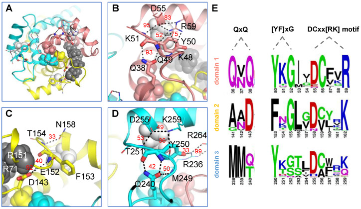
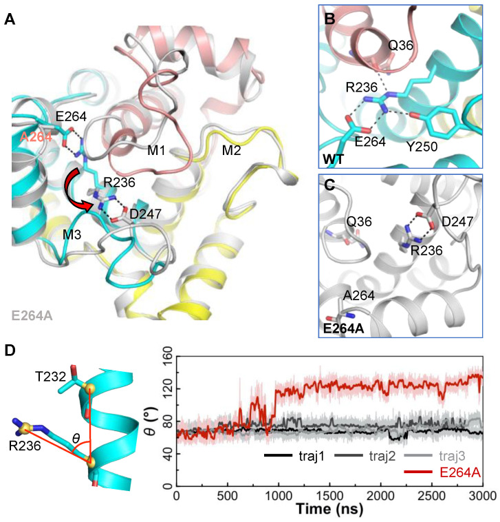
The ADP/ATP carrier (AAC) plays a central role in oxidative metabolism by exchanging ATP and ADP across the inner mitochondrial membrane. Previous experiments have shown the involvement of the matrix loops of AAC in its function, yet potential mechanisms remain largely elusive. One obstacle is the limited information on the structural dynamics of the matrix loops. In the current work, unbiased all-atom molecular dynamics (MD) simulations were carried out on c-state wild-type AAC and mutants. Our results reveal that: (1) two ends of a matrix loop are tethered through interactions between the residue of triplet 38 (Q38, D143 and Q240) located at the C-end of the odd-numbered helix and residues of the [YF]xG motif located before the N-end of the short matrix helix in the same domain; (2) the initial progression direction of a matrix loop is determined by interactions between the negatively charged residue of the [DE]G motif located at the C-end of the short matrix helix and the capping arginine (R30, R139 and R236) in the previous domain; (3) the two chemically similar residues D and E in the highly conserved [DE]G motif are actually quite different; (4) the N-end of the M3 loop is clamped by the [DE]G motif and the capping arginine of domain 2 from the two sides, which strengthens interactions between domain 2 and domain 3; and (5) a highly asymmetric stable core exists within domains 2 and 3 at the m-gate level. Moreover, our results help explain almost all extremely conserved residues within the matrix loops of the ADP/ATP carriers from a structural point of view. Taken together, the current work highlights asymmetry in the three matrix loops and implies a close relationship between asymmetry and ADP/ATP transport.

Keywords: ADP/ATP carrier (AAC); MCF motif; loops; mitochondrial carrier family (MCF); molecular dynamics simulation; transporters.


Conflict of interest statement

The authors declare that they have no conflict of interest with the contents of this article.

Figures

Figure 1
Figure 1
The matrix loops in AAC and conserved MCF motifs nearby. (A) Schematic diagram of the tripartite structure of AAC. Each domain is shown in a different color (domain 1—salmon; domain 2—yellow; domain 3—cyan). The [YF]xG and DCxx[RK] motifs and triplet 38 (Q38, D143 and Q240) within the Px[DE]xx[KR]xRxQxQ motif are highlighted in gray. (B) Sequence alignment of the three domains from triplet 27 (conserved kink prolines) to triplet 65 (the G of the [DE]G motif at the C-end of short matrix helices h12, h34 and h56), with residues numbered based on bovine AAC1. The motif residues mentioned above are highlighted in gray characters. (C) The tertiary structure of AAC in the c-state. The left panel represents a cross-section of AAC and solvent, with the interior of AAC shown in gray and the solvent shown in blue. Cα atoms of the motifs mentioned above are highlighted by small black balls, and residues in the hydrophobic plug (V37, A142 and M239) at the C-end of odd-numbered helices are shown as spheres.
Figure 2
Figure 2
Time evolutions of the intra-domain H-bonds near the matrix side of AAC. (A) The intra-loop H-bonds. The H-bonds formed between triplet 38 residues (Q38, D143 and Q240) and the first residues within the [YF]xG motif (Y50, F153 and Y250) are highlighted with red boxes. (B) The intra-domain H-bonds formed between matrix helices and matrix loops. The H-bonds in domains 1, 2 and 3 are shown in salmon, yellow and blue, respectively. Results shown in this figure were calculated based on traj-1, and results of traj-2 and traj-3 are provided in the Supplementary Material.
Figure 3
Figure 3
Intra-domain interactions between matrix loops and conserved MCF motifs in AAC. (A) Overall electrostatic interactions between matrix loops and conserved MCF motifs from the matrix side view. The conserved cysteine residues within the matrix helices, the R30:R71:R151 stacking structure and the hydrophobic plug are shown in white, black and colored spheres, respectively. Detailed interactions in domain 1, domain 2 and domain 3 are shown in (BD), respectively. Electrostatic interactions are represented by black dashed lines, with the occupancies shown in red numbers. (E) Sequence logos of the QxQ motif, [YF]xG motif and DCxx[RK] motif in orthologs of AAC. The logos were generated by 43 sequences of various AAC subtypes from a total of 24 species (all sequences have been reviewed in UniProt). The residues are numbered based on bovine AAC1. In (AD), the structures of domains 1, 2 and 3 are shown in salmon, yellow and blue, respectively.
Figure 4
Figure 4
The determinants of the initial progression direction of the matrix loops of AAC. (A) Superimposed structures of the three domains to highlight the different progression directions of the three matrix loops. Residues of triplet 38 are presented in stick mode. Detailed interactions in domain 1, domain 2 and domain3 are shown in (BD), respectively. Residues that cause steric hindrance (B) or form a stacking structure (C) are also shown as transparent spheres. In (AD), the structures of domains 1, 2 and 3 are shown in salmon, yellow and blue, respectively.
Figure 5
Figure 5
Impact of the E264A mutation on the structure of AAC. (A) Superimposed structures of the wild-type AAC (in color) and E264A-AAC mutant (in white) at the end of the 3 μs simulations. The flip of the R236 side chain from wild-type conformation to the E264A mutant conformation is highlighted by a red arrow. (B,C) The detailed electrostatic interactions involving R236 in the wild-type AAC and E264A-AAC mutant. These interactions are shown by black dash lines. (D) Time evolutions of the side-chain orientation of R236 (θ) during the simulations of the wild-type AAC and E264A-AAC mutant. Here, θ is defined as the angle between the Cζ atom of R236, the Cα atom of R236 and the Cα atom of T232 (R236Cζ- R236Cα- T232Cα). In (A,B), the structures of domains 1, 2 and 3 of wild-type AAC are shown in salmon, yellow and blue, respectively.
Figure 6
Figure 6
Structural changes in the simulations of E264D-AAC and D167E-AAC mutants. (A) Salt bridges between R236 and E/D264. (B) Time evolutions of minimum distances between residues 236 and 264 in the wild-type AAC (dark lines) and E264D-AAC mutant (red line). (C) The N-end of the M3 loop forms interactions with D/E167 and R137. (D) Time evolutions of minimum distances between residue 167 and the M3 loop in wild-type (dark lines) and D167E mutant AACs (red line). In (A,C), the structures of domains 1, 2 and 3 are shown in salmon, yellow and blue, respectively.
Figure 7
Figure 7
Frequencies of E in the [DE]G motifs of the three homologous domains in different groups of human MCs. Of a total of 53 human mitochondrial carriers, 46 were used for the calculation. Those carriers that are reported to localize outside the IMM (MTCH1, MTCH2, SLC25A46 and PM34) and those having an unexpected charged residue in the second position of the [DE]G motif (SLC25A47, SLC25A51 and SLC25A52) were excluded from the calculation. Please refer to Supplemental Table S1 for detailed information on the members of each group and the first residues of the [DE]G motifs in the three homologous domains of each carrier.
Figure 8
Figure 8
Interactions between a matrix loop and the conserved MCF motif. Electrostatic interactions are highlighted with blue dash lines. A residue is shown by a red letter if it is involved in the interactions through its backbone atoms.
Figure 9
Figure 9
A highly asymmetric stable core exists in domains 2 and 3 of c-state apo AAC. (A) The interlocked aromatic cluster formed between the [YFW][KR]G motif residues and proline kink residues in domain 2. The side chains of C128 and Q174 are not shown for clarity purposes. (B) The interlocked aromatic cluster and the H3H4H5 network wrap tightly around M238. Occupancies for some bonds are shown in red or white numbers, and for complete data, please refer to our previous work [22]. (C) The matrix view of the positions of M237, M239 and the stable R236:E264 salt bridge. (D) The N-end of M3 is clamped by domain 2 through an exquisite electrostatic network. (E) The c-core residues are asymmetrically distributed. (F) The sequence logo presentation of the c-core residues within their triplets. In (AF), the interlocked aromatic cluster in domain 2 is shown in cyan; the H3-H4-H5 network is shown in blue; MMM is shown in magenta; the R236:E264 salt bridge is shown in green; and the network between domain 2 and the N-end of the M3 loop is shown in light orange. Residues are shown first as sticks (with only polar hydrogen atoms) to highlight the interactions and then in spheres (with all hydrogen atoms) to show the space occupancy.

Similar articles

Cited by

References

    1. Lamarca V., Sanz-Clemente A., Pérez-Pé R., Martínez-Lorenzo M.J., Halaihel N., Muniesa P., Carrodeguas J.A. Two isoforms of PSAP/MTCH1 share two proapoptotic domains and multiple internal signals for import into the mitochondrial outer membrane. Am. J. Physiol. Cell Physiol. 2007;293:C1347–C1361. doi: 10.1152/ajpcell.00431.2006. - DOI - PubMed
    1. Zaltsman Y., Shachnai L., Yivgi-Ohana N., Schwarz M., Maryanovich M., Houtkooper R.H., Vaz F.M., De Leonardis F., Fiermonte G., Palmieri F., et al. MTCH2/MIMP is a major facilitator of tBID recruitment to mitochondria. Nat. Cell Biol. 2010;12:553–562. doi: 10.1038/ncb2057. - DOI - PMC - PubMed
    1. Abrams A.J., Hufnagel R.B., Rebelo A., Zanna C., Patel N., Gonzalez M.A., Campeanu I.J., Griffin L.B., Groenewald S., Strickland A.V., et al. Mutations in SLC25A46, encoding a UGO1-like protein, cause an optic atrophy spectrum disorder. Nat. Genet. 2015;47:926–932. doi: 10.1038/ng.3354. - DOI - PMC - PubMed
    1. Visser W.F., van Roermund C.W.T., Waterham H.R., Wanders R.J.A. Identification of human PMP34 as a peroxisomal ATP transporter. Biochem. Biophys. Res. Commun. 2002;299:494–497. doi: 10.1016/S0006-291X(02)02663-3. - DOI - PubMed
    1. Kim Y.-I., Nam I.-K., Lee D.-K., Bhandari S., Charton L., Kwak S., Lim J.-Y., Hong K., Kim S.-J., Lee J.N., et al. Slc25a17 acts as a peroxisomal coenzyme A transporter and regulates multiorgan development in zebrafish. J. Cell. Physiol. 2020;235:151–165. doi: 10.1002/jcp.28954. - DOI - PubMed

MeSH terms

LinkOut - more resources