Review
. 2022 Sep 17;23(18):10871.
doi: 10.3390/ijms231810871.

How "Neuronal" Are Human Skin Mast Cells?

Affiliations
Review

How "Neuronal" Are Human Skin Mast Cells?

Magda Babina et al. Int J Mol Sci. .

Abstract

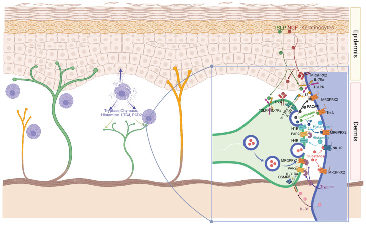
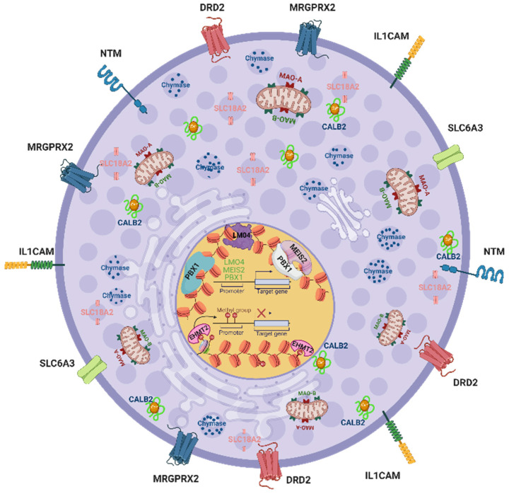
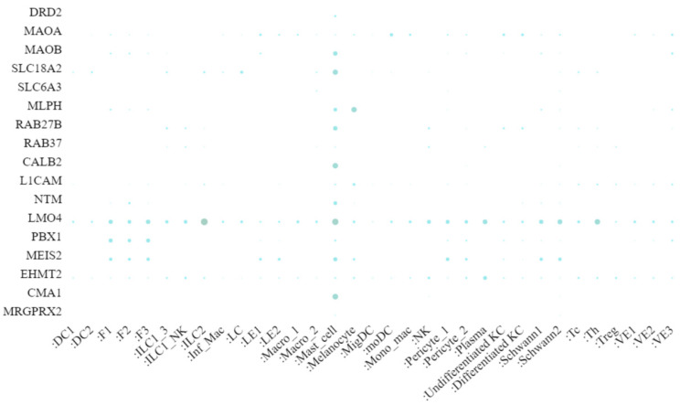
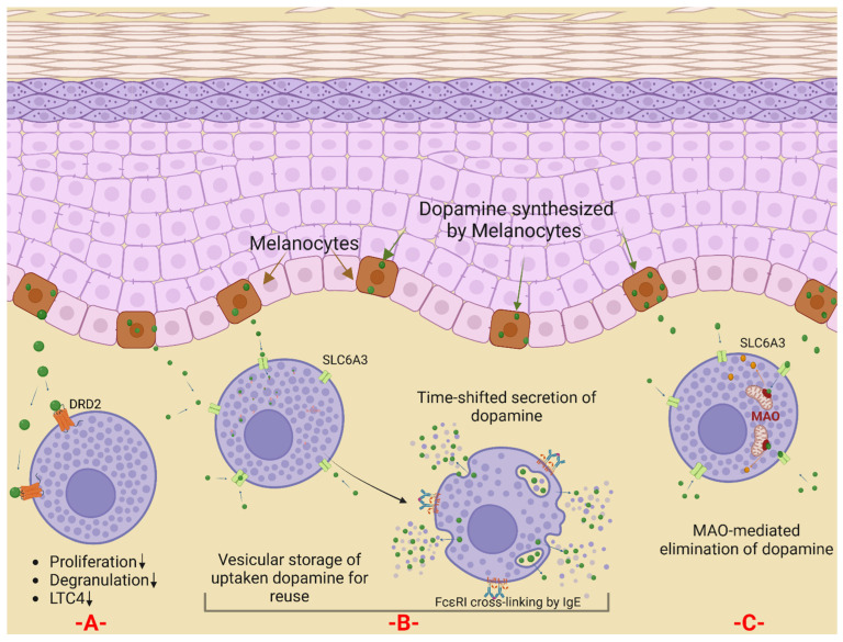
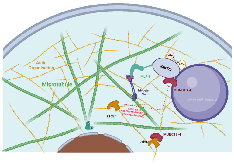
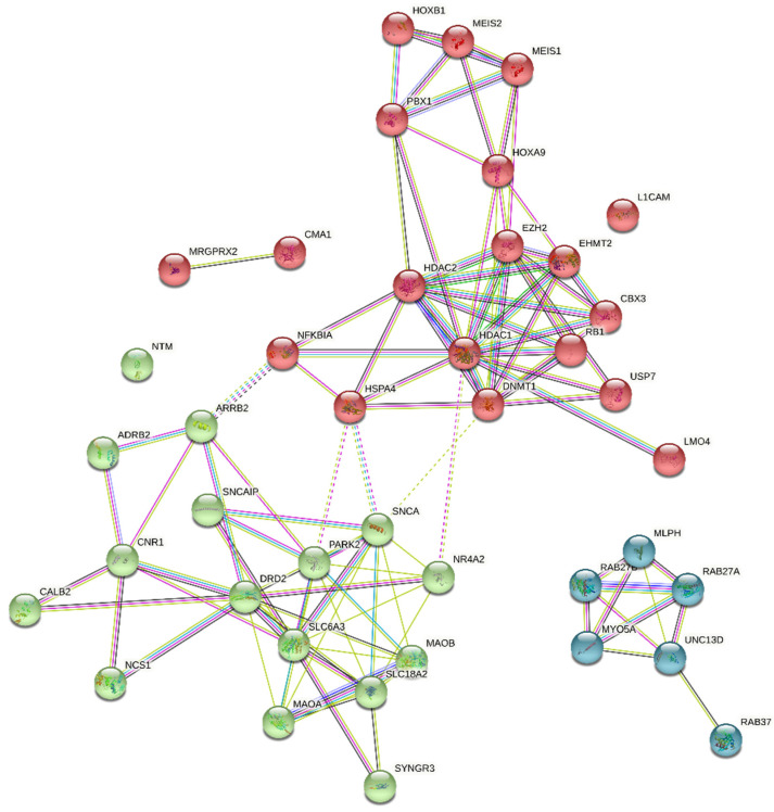
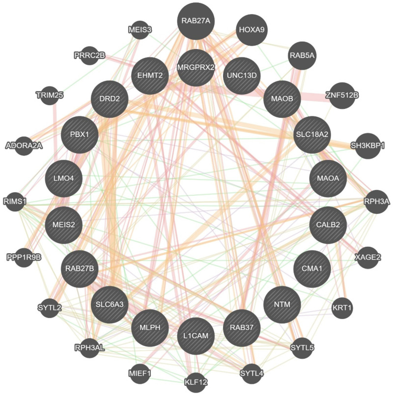
Mast cells are evolutionarily old cells and the principal effectors in allergic responses and inflammation. They are seeded from the yolk sac during embryogenesis or are derived from hematopoietic progenitors and are therefore related to other leukocyte subsets, even though they form a separate clade in the hematopoietic system. Herein, we systematically bundle information from several recent high-throughput endeavors, especially those comparing MCs with other cell types, and combine such information with knowledge on the genes' functions to reveal groups of neuronal markers specifically expressed by MCs. We focus on recent advances made regarding human tissue MCs, but also refer to studies in mice. In broad terms, genes hyper-expressed in MCs, but largely inactive in other myelocytes, can be classified into subcategories such as traffic/lysosomes (MLPH and RAB27B), the dopamine system (MAOB, DRD2, SLC6A3, and SLC18A2), Ca2+-related entities (CALB2), adhesion molecules (L1CAM and NTM) and, as an overall principle, the transcription factors and modulators of transcriptional activity (LMO4, PBX1, MEIS2, and EHMT2). Their function in MCs is generally unknown but may tentatively be deduced by comparison with other systems. MCs share functions with the nervous system, as they express typical neurotransmitters (histamine and serotonin) and a degranulation machinery that shares features with the neuronal apparatus at the synapse. Therefore, selective overlaps are plausible, and they further highlight the uniqueness of MCs within the myeloid system, as well as when compared with basophils. Apart from investigating their functional implications in MCs, a key question is whether their expression in the lineage is due to the specific reactivation of genes normally silenced in leukocytes or whether the genes are not switched off during mastocytic development from early progenitors.

Keywords: adhesion molecules; calbindin; degranulation; dopamine; mast cells; monoamine oxidase B; neurons; skin; solute carriers; transcription factors.


Conflict of interest statement

The authors declare no conflict of interest.

Figures

Figure 1
Figure 1
MCs communicate with sensory nerves in the skin. MCs are frequently situated near neurons, whereby the two are engaged in a close dialogue. Neuropeptides such as SP and cortistatin degranulate MCs, chiefly by activating on MRGPRX2; NK-1R can be contributory in the case of SP. Mediators exocytosed from MCs comprise the pruritogens histamine and tryptase, which act on H1R, H4R, and PAR2 on nerve endings (and potentially Mrgprs, i.e., MRGPRX1) on sensory neurons. In addition, the MC-derived cytokines IL-13 and IL-31 can further prime sensory neurons and induce their growth and sprouting. In the skin, keratinocytes occupy an important role and strongly influence both neurons and MCs, e.g., by provision of NGF and TSLP. See text for further details. For the sake of simplicity, the communication is depicted for one nerve only; however, in reality, different receptor combinations characterize subsets of sensory neurons, whereby distinct entities exclude others. Abbreviations: H1R (Histamine H1 receptor), H4R (Histamine H4 receptor), IL-13 (Interleukin-13), IL-13RA1 (Interleukin-13 receptor alpha 1), IL-31R (Interleukin-31 receptor), IL-7Ra (Interleukin 7 receptor alpha), LTC4 (Leukotriene C4), MRGPRX1 (Mas-related G-protein-coupled receptor member X1), MRGPRX2 (Mas-related G-protein-coupled receptor member X2), NGF (Beta-nerve growth factor), NK-1R (Neurokinin receptor-1), OSMRß (Oncostatin-M-specific receptor subunit beta), PACAP (Pituitary adenylyl cyclase-activating protein), PAR2 (Proteinase-activated receptor 2), PGD2 (Prostaglandin D2), TrkA (Tyrosine kinase receptor A), TSLP (Thymic stromal lymphopoietin), TSLPR (Thymic stromal lymphopoietin receptor), and VIP (Vasoactive intestinal peptide).
Figure 2
Figure 2
The nervous system-related genes covered in this review. A unifying element is their preferential expression in MCs in comparison to other myeloid cells. Depicted is the principal cellular localization of the following entities: DRD2, MAOA, MAOB, SLC18A2, SLC6A3, MLPH, RAB27B, RAB37, CALB2, L1CAM, NTM, LMO4, PBX1, MEIS2, and EHMT2. The genes MC chymase (CMA1) and MRGPRX2, expressed only by MCs and not by other constituents of the body (according to the comprehensive FANTOM5 atlas), have been added for comparison. Abbreviations: CALB2 (Calbindin 2 or Calretinin), DRD2 (Dopamine Receptor D2), EHMT2 (Histone-lysine N-methyltransferase), L1CAM (Neural cell adhesion molecule L1), LMO4 (LIM domain transcription factor 4), MAOA (Amine oxidase (flavin-containing) A), MAOB (Amine oxidase (flavin-containing) B), MEIS2 (Myeloid-Ecotropic Viral Integration Site 1 Homolog 2), MRGPRX2 (Mas-related G-protein-coupled receptor member X2), NTM (Neurotrimin), PBX1 (Pre-B-cell leukemia transcription factor 1), SLC18A2 (Synaptic vesicular amine transporter), and SLC6A3 (Sodium-dependent dopamine transporter).
Figure 3
Figure 3
scRNAseq data of healthy adult skin. The expression of selected genes was extracted from scRNAseq analyses of healthy adult skin and is presented as dot plots; the data are from Reynolds et al. [82]. The color-coded size of the points corresponds with the expression level (the larger and darker, the more highly expressed). (https://cells.ucsc.edu/?ds=healthy-human-skin, accessed last on 1 July 2022) In the associated study [82], the transcriptomes of 200,462 healthy, single skin cells, which were classified into 34 different cell types, were sequenced using the scRNA-seq platform (10× Genomics); the link to the atlas can be found here: https://cells.ucsc.edu/?ds=healthy-human-skin, accessed last on 1 July 2022. Abbreviations: DC, dendritic cell; F, fibroblast; Inf_Mac, inflammatory macrophage; ILC, innate lymphoid cell; KC, keratinocyte; LC, Langerhans cell; LE, lymphatic endothelium; Mono mac, monocyte-derived macrophage; Mig., migratory; MoDC, monocyte-derived dendritic cell; NK, natural killer cell; Tc, cytotoxic T cell; TH, T helper cell; Treg, regulatory T cell; VE, vascular endothelium. The numbers 1, 2, and 3 represent different states or subtypes of cell types.
Figure 4
Figure 4
Hypothetical representation of the connections between dopamine and skin mast cells. Some skin cells, including melanocytes, can produce dopamine under physiologic and/or pathological conditions. (A) By virtue of their DRD2 receptor, skin MCs can respond to dopamine, which, analogously to other MCs, may dampen their proliferation and function. (B) MCs can take up dopamine via SLC6A3. The exogenously provided dopamine can be transported to MC granules and be exocytosed upon appropriate stimulation later. (C) Upon dopamine uptake via SLC6A3, dopamine may be transported to mitochondria to be degraded by MAO-A and/or MAO-B. In this latter scenario, MCs would function as dopamine scavengers.
Figure 5
Figure 5
The granule transport machinery of MCs with involvement of melanophilin and Rab proteins. The intracellular transport of cytoplasmic granules to the MC surface for exocytosis requires a complex motor apparatus connected to the granule membrane, in which a motor protein myosin Va and microtubules work together. In this machinery, melanophilin (MLPH) directly connects myosin Va with Rab proteins located on the granule membrane. Subsequent docking to the plasma membrane is carried out by the protein unc-13 homolog D, which in turn can be blocked by RAB37. See main text for further details. Abbreviations: MLPH (melanophilin), MUNC13-4 (Protein unc-13 homolog D), RAB27A (Ras-related protein Rab-27A), RAB27B (Ras-related protein Rab-27B), and RAB37 (Ras-related protein Rab37).
Figure 6
Figure 6
Protein–protein interaction network from the String Database. To predict the functional interactions of selected neuronal genes with other genes transcribed in MCs, a network analysis was performed using the STRING database (https://string-db.org) [242]. Up to the fourth step, additional nodes were added to the basal model until interconnectivity was achieved. The final network is displayed as a specified number (kmeans = 3) of clusters according to its topological properties, with each cluster highlighted in a different color.
Figure 7
Figure 7
Protein–protein interaction network based on the GeneMANIA web server. To find putative interactions of selected neuronal genes with other partners, network analysis was performed using the GeneMANIA web server [246]. Each gene is a node and evidence for interactions between protein pairs are edges. The thickness of the edges indicates the strength of the interaction, the color of the edges indicates the type of interaction, and the size of the nodes distinguishes the hub gene (inner circles) from the predicted genes (outer circles). Several of the predicted genes are transcribed in MCs (e.g., ADORA2A, SYTL4, KRT1, and MIEF1, and some at substantial levels, e.g., SYTL2).

Similar articles

Cited by

References

    1. Minai-Fleminger Y., Levi-Schaffer F. Mast cells and eosinophils: The two key effector cells in allergic inflammation. Inflamm. Res. 2009;58:631–638. doi: 10.1007/s00011-009-0042-6. - DOI - PubMed
    1. Metcalfe D.D., Peavy R.D., Gilfillan A.M. Mechanisms of mast cell signaling in anaphylaxis. J. Allergy Clin. Immunol. 2009;124:639–646; quiz 647–678. doi: 10.1016/j.jaci.2009.08.035. - DOI - PMC - PubMed
    1. Gilfillan A.M., Beaven M.A. Regulation of mast cell responses in health and disease. Crit. Rev. Immunol. 2011;31:475–529. doi: 10.1615/CritRevImmunol.v31.i6.30. - DOI - PMC - PubMed
    1. Galli S.J., Tsai M. IgE and mast cells in allergic disease. Nat. Med. 2012;18:693–704. doi: 10.1038/nm.2755. - DOI - PMC - PubMed
    1. Kuhn H., Kolkhir P., Babina M., Dull M., Frischbutter S., Fok J.S., Jiao Q., Metz M., Scheffel J., Wolf K., et al. Mas-related G protein-coupled receptor X2 and its activators in dermatologic allergies. J. Allergy Clin. Immunol. 2021;147:456–469. doi: 10.1016/j.jaci.2020.08.027. - DOI - PubMed

MeSH terms

LinkOut - more resources