Review
. 2022 Sep 17;23(18):10900.
doi: 10.3390/ijms231810900.

The Controversial Role of LPS in Platelet Activation In Vitro

Affiliations
Review

The Controversial Role of LPS in Platelet Activation In Vitro

Luca Galgano et al. Int J Mol Sci. .

Abstract

Circulating platelets are responsible for hemostasis and thrombosis but are also primary sensors of pathogens and are involved in innate immunity, inflammation, and sepsis. Sepsis is commonly caused by an exaggerated immune response to bacterial, viral, and fungal infections, and leads to severe thrombotic complications. Among others, the endotoxin lipopolysaccharide (LPS) found in the outer membrane of Gram-negative bacteria is the most common trigger of sepsis. Since the discovery of the expression of the LPS receptor TLR4 in platelets, several studies have investigated the ability of LPS to induce platelet activation and to contribute to a prothrombotic phenotype, per se or in combination with plasma proteins and platelet agonists. This issue, however, is still controversial, as different sources, purity, and concentrations of LPS, different platelet-purification protocols, and different methods of analysis have been used in the past two decades, giving contradictory results. This review summarizes and critically analyzes past and recent publications about LPS-induced platelet activation in vitro. A methodological section illustrates the principal platelet preparation protocols and significant differences. The ability of various sources of LPS to elicit platelet activation in terms of aggregation, granule secretion, cytokine release, ROS production, and interaction with leukocytes and NET formation is discussed.

Keywords: E. coli; LPS; TLR4; platelets; sepsis; thromboinflammation.


Conflict of interest statement

The authors declare no conflict of interest.

Figures
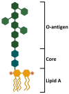
Figure 1
Figure 1
Structure of E. coli LPS. LPS is a complex molecule made of lipids and sugars. The structure is divided into three different domains. The lipid A moiety is localized into the outer membrane of the Gram-negative bacteria and is responsible for TLR4 interaction through the phosphate groups and the acyl chains. The central core is composed of oligosaccharide and links the lipid A moiety with the O-antigen. The O-antigen is made of polysaccharides and can reach over 100 repeats of sugars. The complexity and the high number of repeats allow a more stringent barrier against antibody recognition. Furthermore, the O-antigen allows differentiating bacterial species and serotypes [21,22].
Figure 2
Figure 2
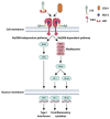
LPS–TLR4 signaling cascades in nucleated cells. LPS-binding protein (LBP) binds to LPS, inducing the exposure of the lipid A moiety and acting as a lipid-transfer protein. LBP drives the interaction of LPS with CD14, a glycosylphosphatidyl inositol-anchored glycoprotein. CD14 acts as a monocyte membrane coreceptor, but it can be released as a soluble protein. The interaction with CD14 allows the transfer of LPS to myeloid differentiation factor 2 (MD-2), a protein that interacts and cooperates with TLR4, activating the signal transduction. MyD88-dependent pathway involves MyD88, TIRAP, IRAK4, and IRAK 1/2, constituting the Myddosome. It activates TRAF6, which in turn activates two transcription factors (AP-1 and NF-κB) that allow the expression of proinflammatory cytokines. MyD88-independent pathway does not involve MyD88, but activates TRAF3, which in turn activates IRF3, another transcription factor, involved in type I interferon expression [21,22,27,29].
Figure 3
Figure 3
LPS–TLR4 signaling cascades in platelets. LPS initiates the MyD88-dependent signaling cascade, which in turn activates many downstream effectors (Erk1/2, p38 MAPK, cPLA2, Akt, JNK, NF-κB, and p47phox), leading to or potentiating platelet aggregation, granule secretion, IL-1β synthesis and release, and ROS production [10,12,13,14,15,16,17,19,20,21,44,45,50]. The role of MyD88-independent pathway is still unclear.

References

    1. Thon J.N., Italiano J.E. Platelets: Production, Morphology and Ultrastructure. In: Gresele P., Born G., Patrono C., Page C., editors. Antiplatelet Agents. Springer; Berlin/Heidelberg, Germany: 2012. pp. 3–22. (Handbook of Experimental Pharmacology Series No. 210). - DOI - PubMed
    1. Krishnegowda M., Rajashekaraiah V. Platelet disorders: An overview. Blood Coagul. Fibrinolysis. 2015;26:479–491. doi: 10.1097/01.mbc.0000469521.23628.2d. - DOI - PubMed
    1. Holinstat M. Normal platelet function. Cancer Metastasis Rev. 2017;36:195–198. doi: 10.1007/s10555-017-9677-x. - DOI - PMC - PubMed
    1. Jenne C.N., Kubes P. Platelets in inflammation and infection. Platelets. 2015;26:286–292. doi: 10.3109/09537104.2015.1010441. - DOI - PubMed
    1. Zucoloto A.Z., Jenne C.N. Platelet-Neutrophil Interplay: Insights into Neutrophil Extracellular Trap (NET)-Driven Coagulation in Infection. Front. Cardiovasc. Med. 2019;6:85. doi: 10.3389/fcvm.2019.00085. - DOI - PMC - PubMed